Clinical Reference for Dental Professionals

Which CBCT Findings Change the Implant Treatment Plan?

Identify which CBCT findings should alter implant position, dimensions, site development, staging, or referral decisions.

πŸŽ₯ CoDL Expert Perspective (Coming Soon)

Watch a short clinician discussion on how experienced dentists use CBCT to modify implant position, dimensions, site development, and referral decisions.

🧭 Clinical Decision Snapshot

The relevant question is not simply "Is bone present?" but "Can the anatomy safely support the planned implant and definitive restoration at this site?"

CBCT should be prescribed when additional three-dimensional information is expected to change diagnosis or treatment planning. It should not be ordered routinely; it should answer a defined clinical question about the implant site, bone, anatomy, pathology, or restorative position.

CBCT can materially affect implant planning and implant-size selection, while incidental findings may require additional assessment or treatment.

Key Clinical Takeaways
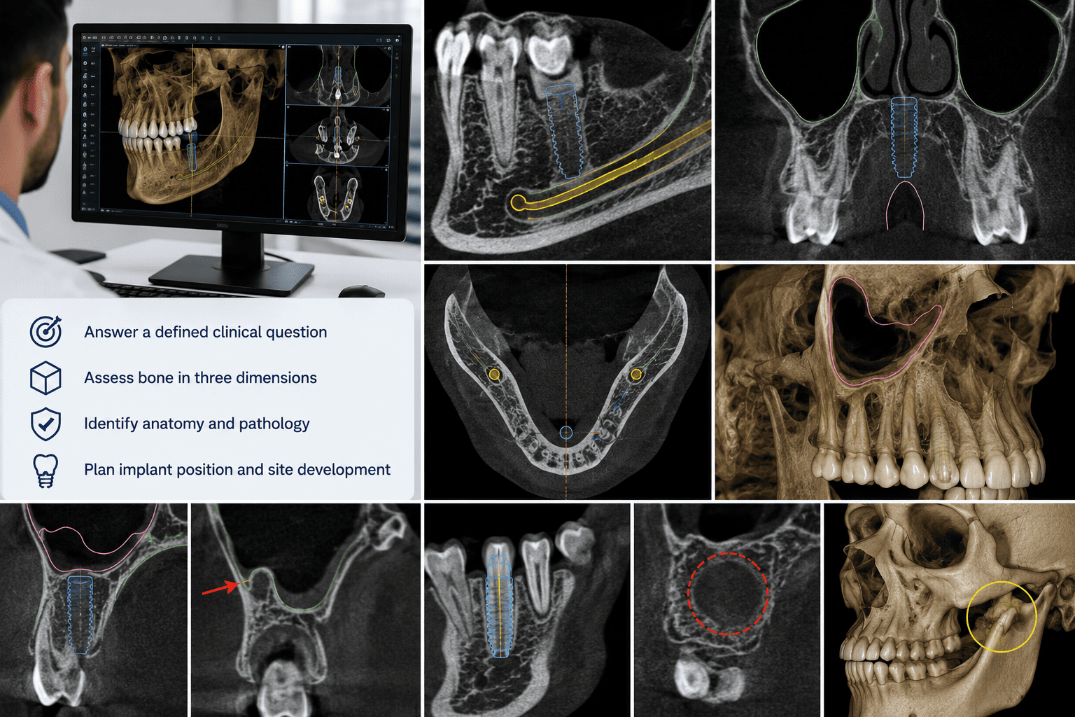
A dentist reviewing a 3D CBCT reconstruction and multiplanar views of an implant site on a workstation monitor, alongside a checklist for CBCT interpretation: answer a defined clinical question, assess bone in three dimensions, identify anatomy and pathology, and plan implant position and site development. Surrounding panels show CBCT cross-sections of implant sites near the inferior alveolar canal, maxillary sinus, and adjacent roots, plus a 3D skull reconstruction. Demonstrates the range of CBCT findings that influence implant surgical planning, site development, and referral decisions.
Enlarge

πŸ” Clinical Assessment

Evaluate bone, anatomy, pathology, and incidental findings that affect implant placement and restoration.

What CBCT Findings Should Be Evaluated at the Implant Site?

CBCT interpretation for implant planning should focus on findings that affect implant position, dimensions, site development, staging, or referral at the proposed implant site.

CBCT Assessment Workflow
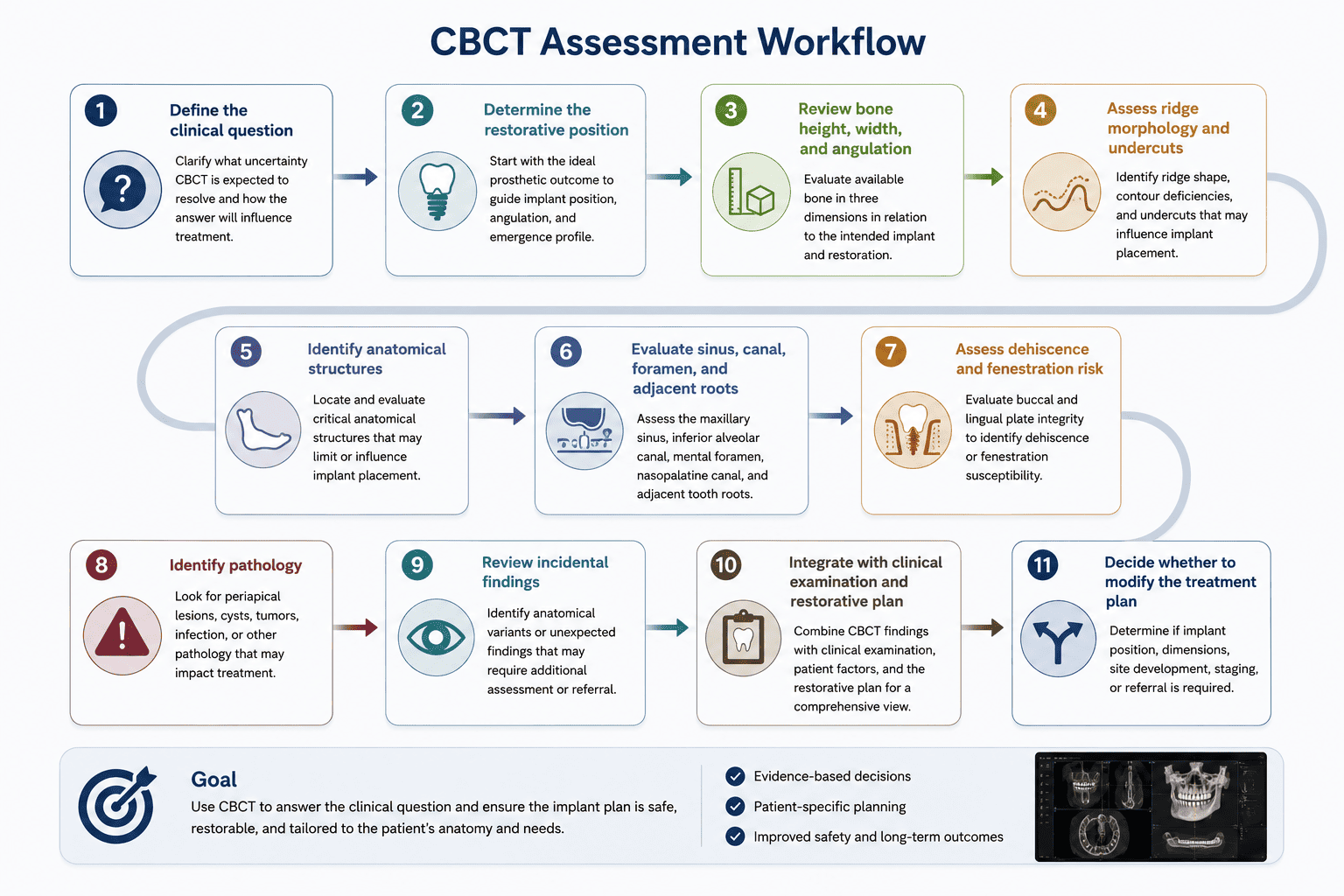
An 11-step CBCT Assessment Workflow diagram: define the clinical question, determine the restorative position, review bone height, width and angulation, assess ridge morphology and undercuts, identify anatomical structures, evaluate sinus, canal, foramen and adjacent roots, assess dehiscence and fenestration risk, identify pathology, review incidental findings, integrate with the clinical examination and restorative plan, and decide whether to modify the treatment plan. Goal: use CBCT to answer the clinical question and ensure the implant plan is safe, restorable, and tailored to the patient's anatomy and needs.
Enlarge
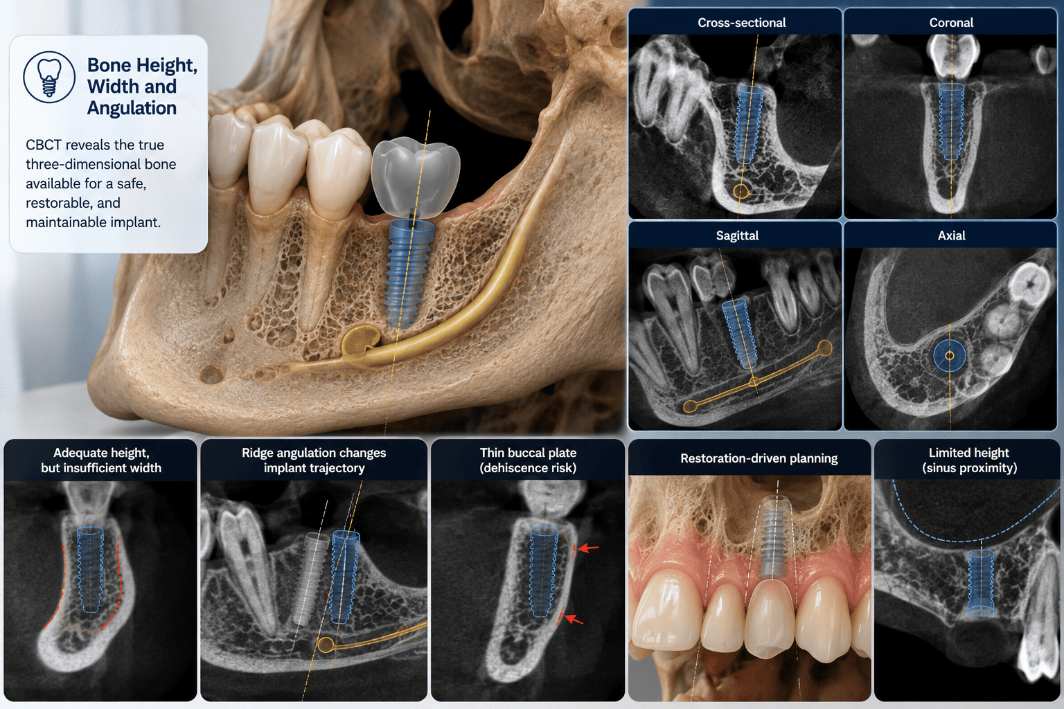
CBCT multiplanar views (cross-sectional, coronal, sagittal, and axial) of a mandibular implant site with bone height, width, and angulation measured against the planned implant, alongside comparison panels labelled adequate height but insufficient width, ridge angulation changing the implant trajectory, thin buccal plate with dehiscence risk, restoration-driven planning, and limited height from sinus proximity. Demonstrates three-dimensional evaluation of bone availability before determining implant position and dimensions.
Enlarge
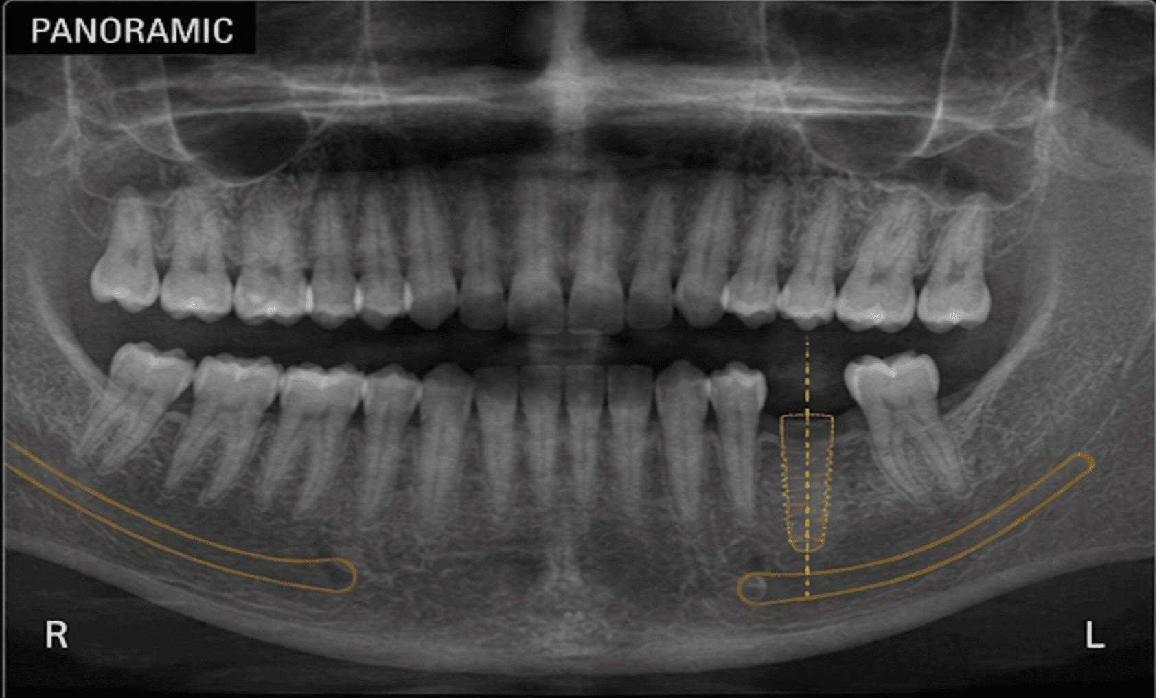
Panoramic radiograph (OPG) showing the full mandibular and maxillary arches, used for initial anatomical screening, including the outlined mandibular canal and a planned implant position at a lower left molar site ahead of targeted CBCT assessment. Demonstrates the role of a panoramic radiograph in global anatomical screening before three-dimensional bone assessment.
Enlarge
Bone Height, Width, and Angulation

Clinical Assessment

Evaluate:

  • Bone height at the proposed implant site
  • Bone width at the crest and along the implant length
  • Ridge angulation relative to the planned implant axis
  • Buccal and lingual plate thickness
  • Relationship to the intended restoration and emergence profile

Clinical Reasoning

Review multiplanar and cross-sectional views before determining implant position, rather than relying on a single linear bone-height measurement.

A common pitfall is measuring bone height without considering ridge angulation, buccal plate thickness, or the intended restorative position.

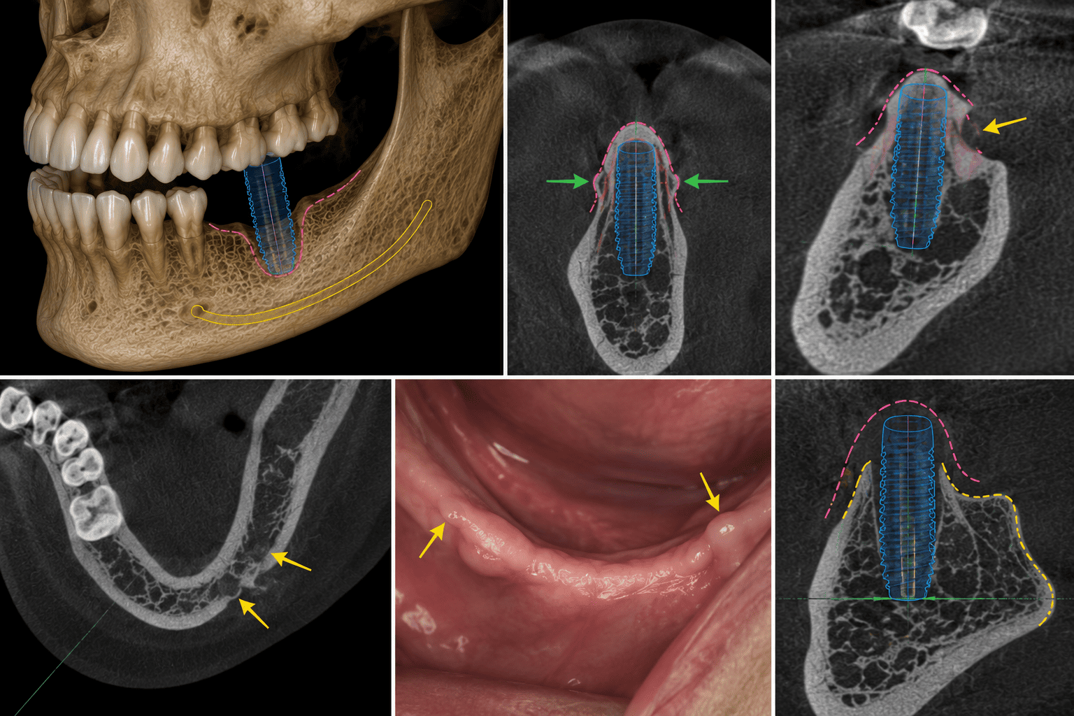
3D CBCT reconstruction and cross-sectional views of a mandibular implant showing a buccal ridge concavity and thin buccal plate outlined around the implant, with green arrows marking the narrowest points and an intraoral photograph showing corresponding mucosal thinning over the site. Demonstrates how ridge morphology and undercuts are assessed for their effect on implant angulation and buccal plate integrity.
Enlarge
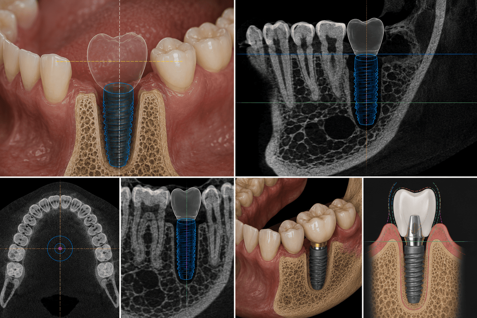
CBCT implant planning views showing a translucent digital restorative crown positioned above the implant across intraoral, sagittal, axial, and coronal slices, with the crown's emergence profile guiding the implant position beneath it. Demonstrates restoration-driven, prosthetically guided CBCT treatment planning.
Enlarge
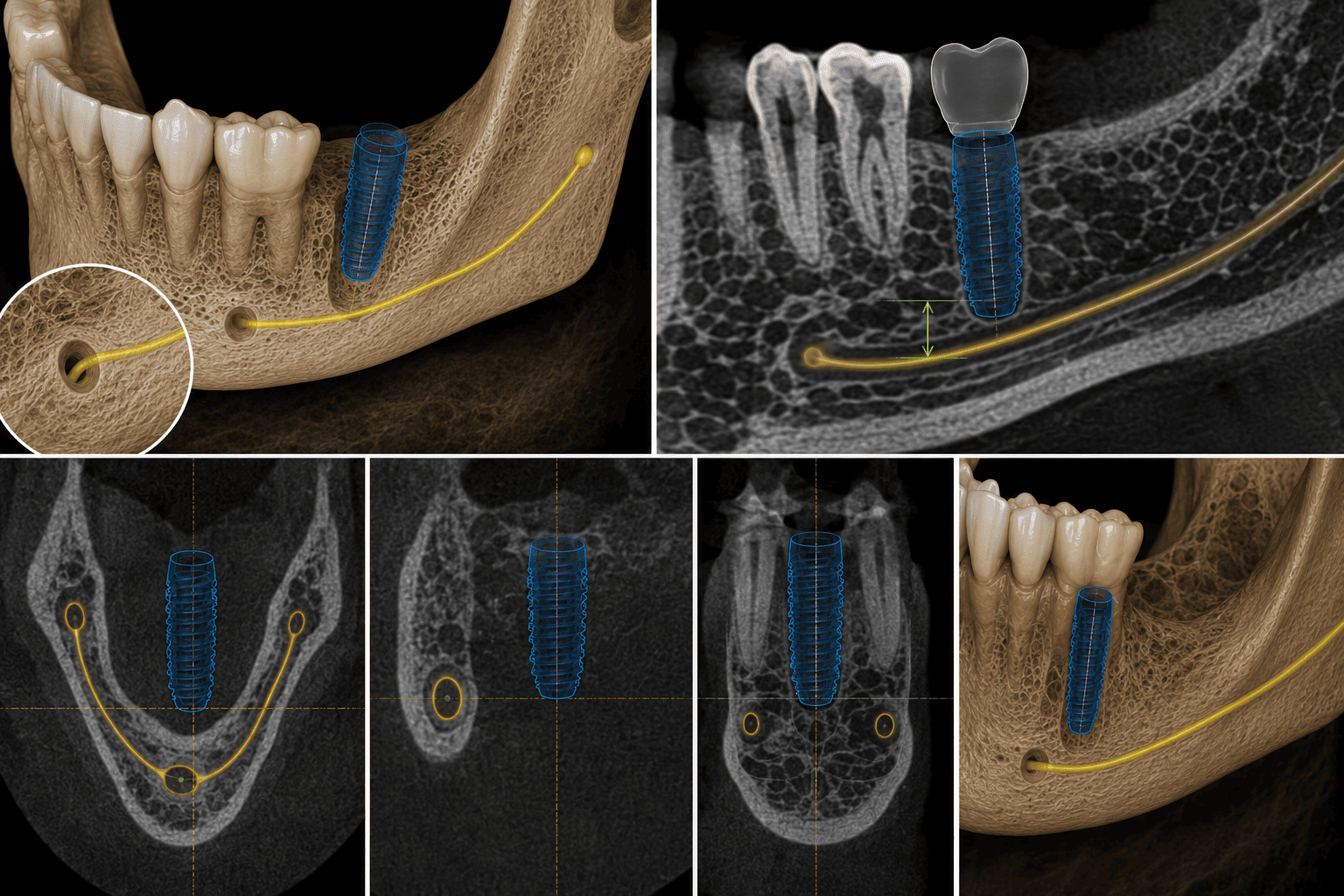
3D CBCT reconstruction and cross-sectional views tracing the inferior alveolar canal from the mandibular foramen forward, with the measured vertical distance from the canal to the planned implant apex and axial slices showing the canal's position relative to adjacent implant sites. Demonstrates identification of the inferior alveolar canal and mental foramen to maintain a safe surgical margin.
Enlarge
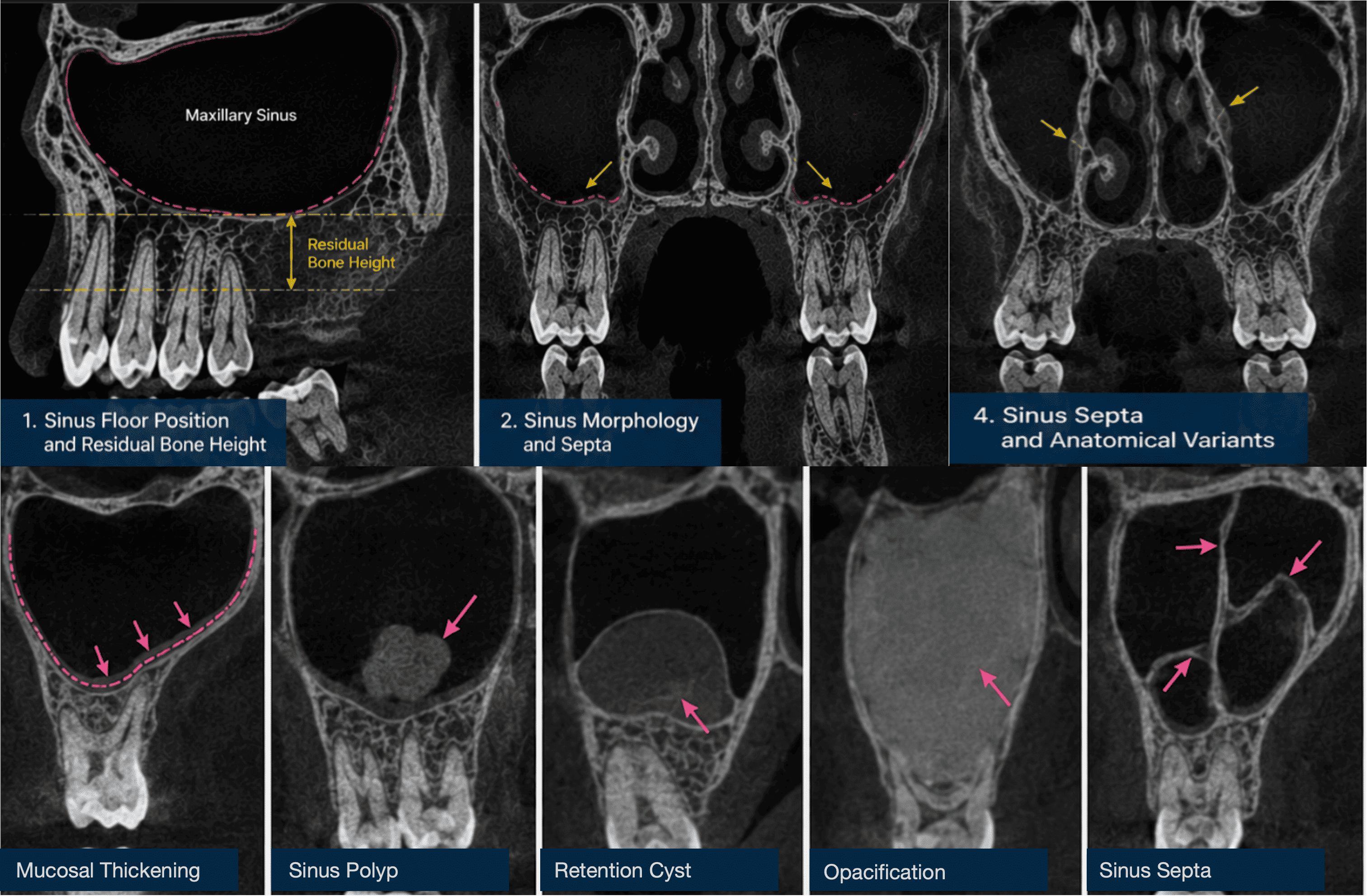
Labelled CBCT coronal views of the maxillary sinus showing sinus floor position and residual bone height, sinus morphology and septa, and anatomical variants, alongside close-up panels illustrating mucosal thickening, a sinus polyp, a retention cyst, opacification, and sinus septa. Demonstrates assessment of sinus floor position, residual bone height, and sinus pathology before posterior maxillary implant planning.
Enlarge
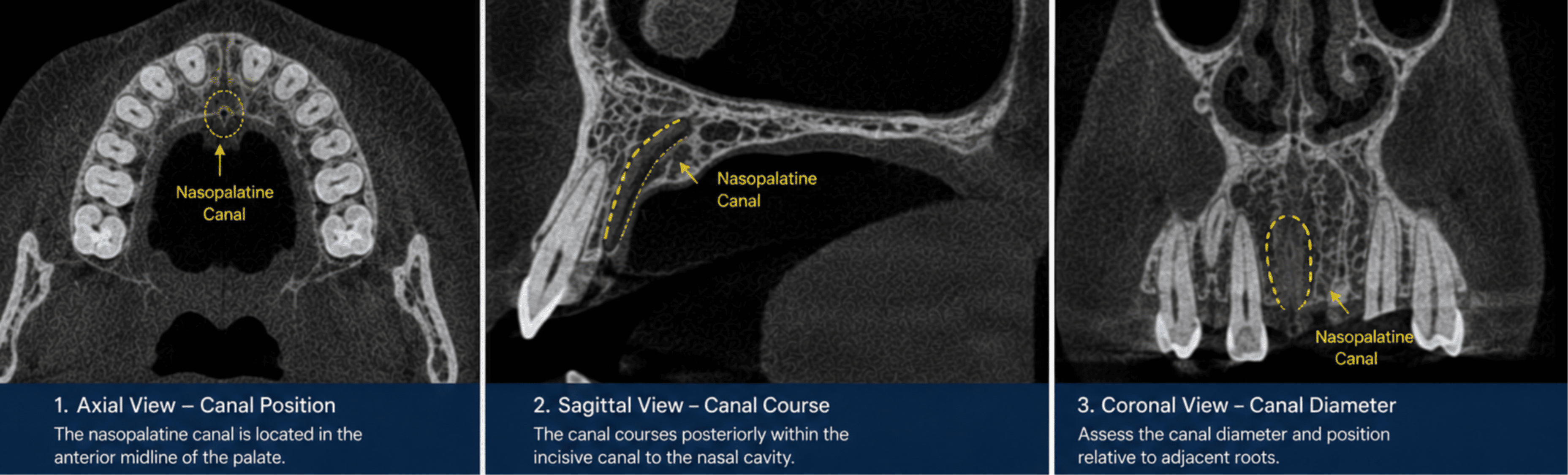
Labelled axial, sagittal, and coronal CBCT views of the anterior maxilla showing the nasopalatine canal in the midline of the palate, its posterior course through the incisive canal toward the nasal cavity, and its diameter relative to adjacent roots. Demonstrates assessment of nasopalatine canal position and risk of encroachment during anterior maxillary implant planning.
Enlarge
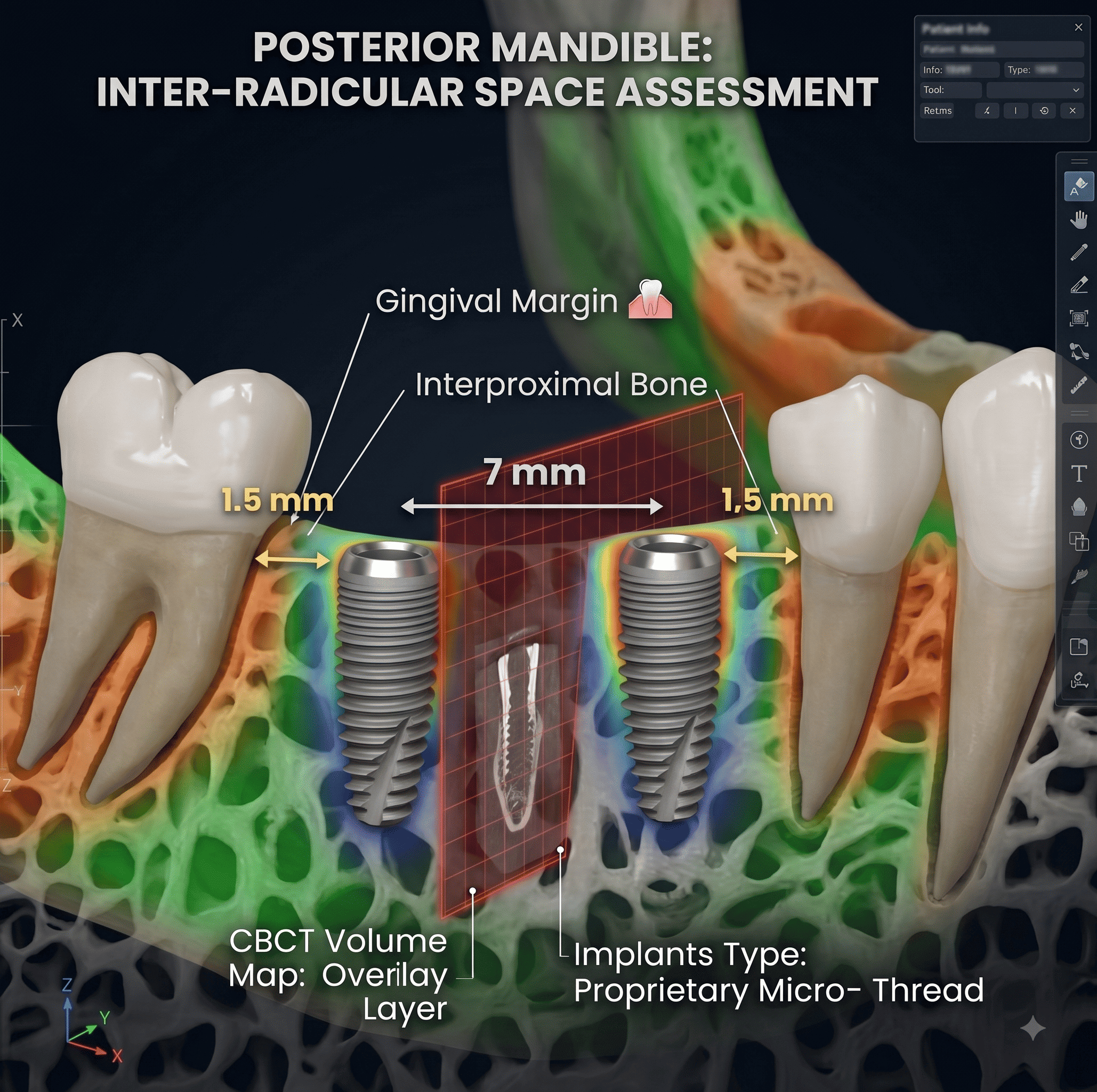
3D CBCT reconstruction of the posterior mandible showing two implants placed between adjacent tooth roots, with the inter-radicular space (7 mm) and the 1.5 mm clearance from each implant to the neighbouring root's gingival margin labelled. Demonstrates assessment of adjacent root proximity and inter-radicular space before finalising implant position.
Enlarge
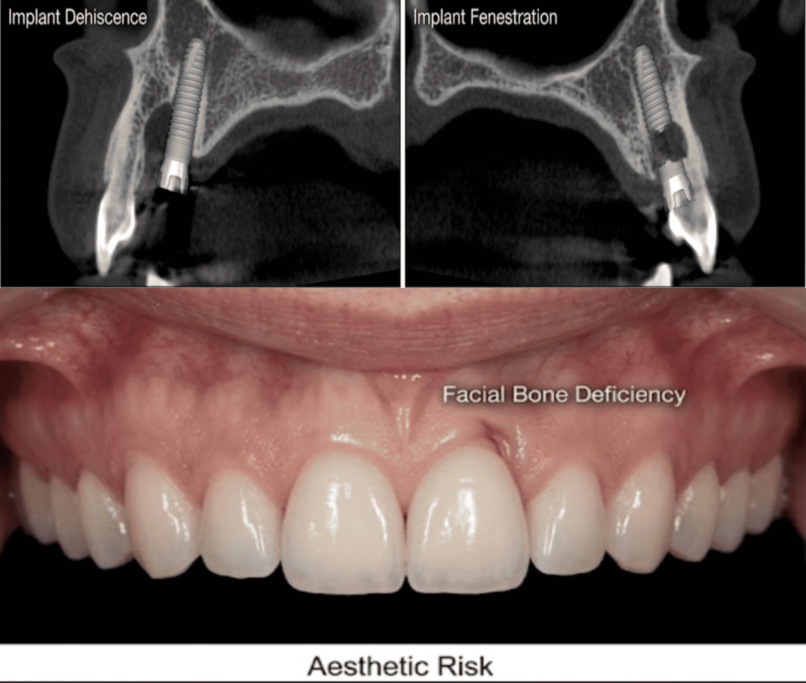
Side-by-side CBCT cross-sections labelled implant dehiscence, showing an implant with exposed buccal threads above the bone crest, and implant fenestration, showing an implant perforating the buccal plate mid-body, alongside an intraoral photograph of anterior facial bone deficiency and associated aesthetic risk. Demonstrates assessment of dehiscence and fenestration risk before implant placement.
Enlarge
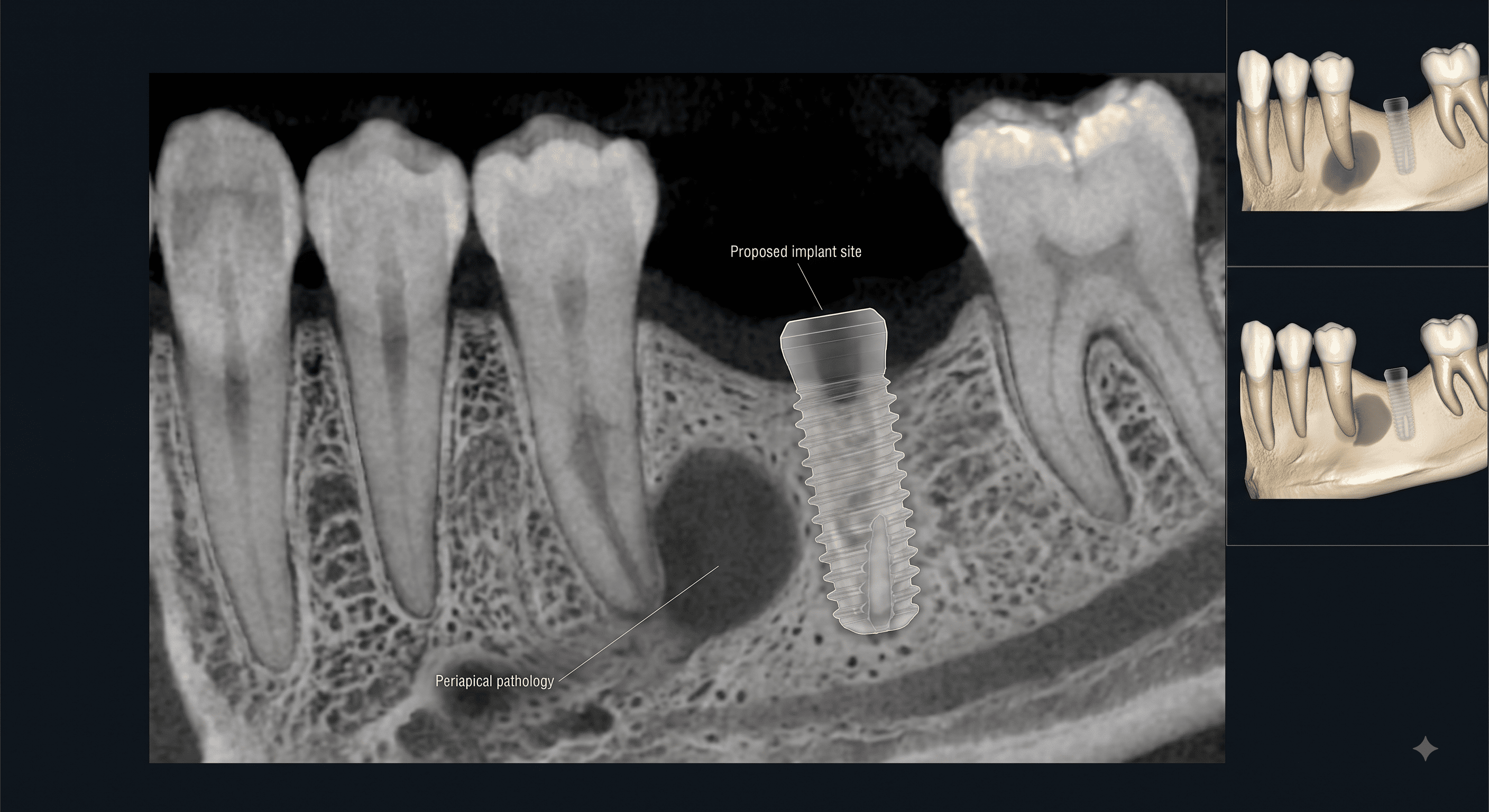
CBCT cross-sectional view labelled periapical pathology, showing a rounded radiolucent lesion at the apex of an adjacent tooth directly beside a proposed implant site, with 3D reconstruction insets comparing the lesion's proximity to the planned implant before and after treatment. Demonstrates identification of existing pathology that may require treatment before implant placement.
Enlarge
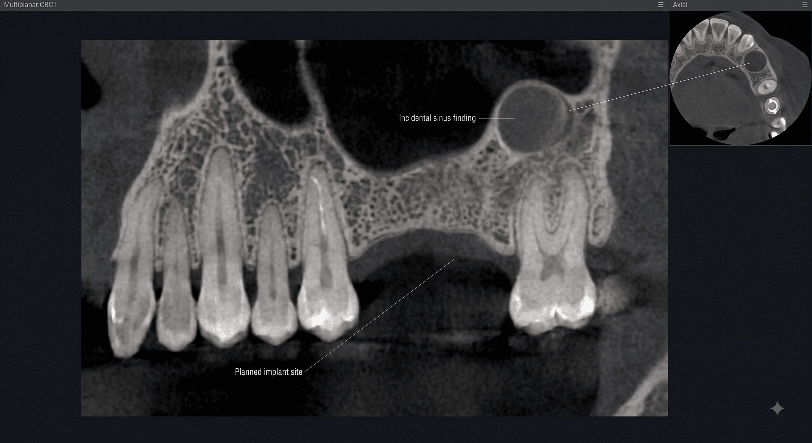
Multiplanar CBCT view labelled with a planned implant site in the posterior maxilla and, well away from it, an incidental sinus finding highlighted on both the coronal slice and an inset axial view. Demonstrates how CBCT may reveal incidental findings unrelated to the original implant planning question.
Enlarge
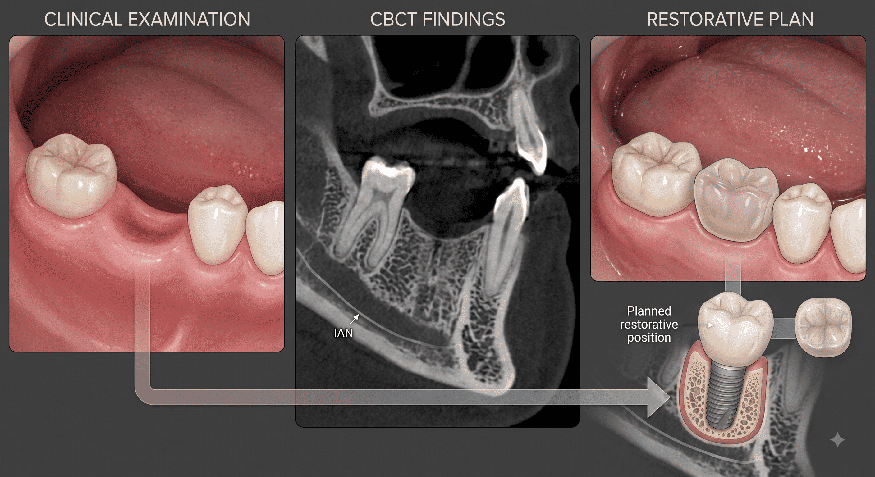
Three-panel illustration labelled Clinical Examination, CBCT Findings, and Restorative Plan, showing an edentulous ridge, a CBCT slice with the inferior alveolar nerve traced beneath adjacent roots, and the planned restorative crown position, connected by an arrow into one integrated implant placement outcome. Demonstrates how CBCT findings are interpreted together with the clinical examination and restorative plan rather than in isolation.
Enlarge

Clinical Pearl

CBCT findings should change the plan whenever they compromise safety, suitability, or maintainability.

CBCT findings should change the treatment plan when they reveal that the original implant position, dimensions, or approach would compromise safety, restorative suitability, or long-term maintainability.

βš–οΈ Clinical Decision-Making

CBCT findings should change the plan whenever they reveal risk to safety, restorative suitability, or long-term maintainability.

How Should CBCT Findings Change the Implant Treatment Plan?

CBCT findings should change the treatment plan when they reveal that the original implant position, dimensions, or approach would compromise safety, restorative suitability, or long-term maintainability at the implant site.

Clinical Decision Framework

How do CBCT findings become one implant treatment recommendation?

Findings Support the Original Plan

Typical Findings

Typical findings include:

  • Adequate bone height, width, and angulation
  • Favourable ridge morphology
  • Safe distance from anatomical structures
  • No significant pathology
  • No incidental findings requiring action

Clinical Reasoning

When these findings converge, the case can proceed with the planned treatment as originally designed.
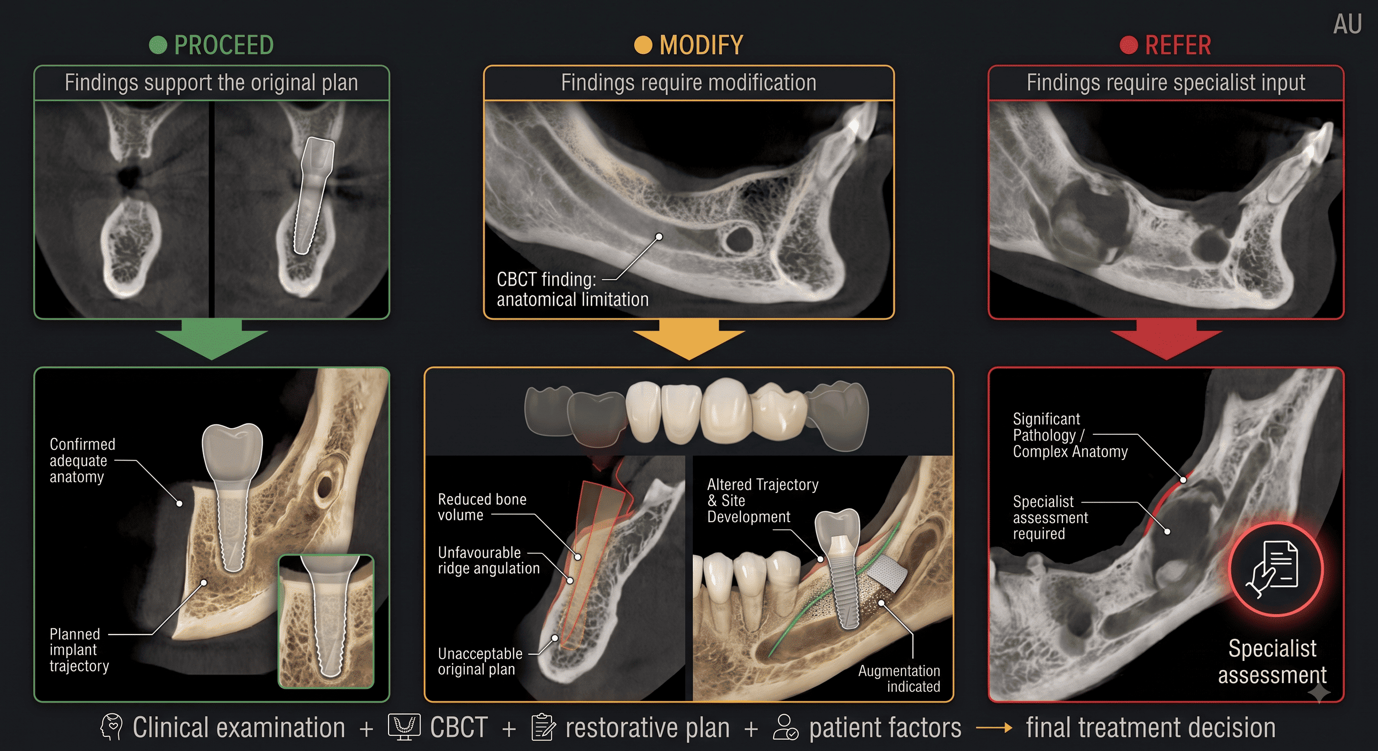
Three-column clinical decision flowchart: PROCEED, where findings support the original plan with confirmed adequate anatomy and the planned implant trajectory; MODIFY, where a CBCT finding of anatomical limitation such as reduced bone volume or unfavourable ridge angulation makes the original plan unacceptable and leads to an altered trajectory, site development, and augmentation; and REFER, where significant pathology or complex anatomy requires specialist assessment. Each pathway combines clinical examination, CBCT, the restorative plan, and patient factors into the final treatment decision.
Enlarge

Clinical Pearl

The strongest implant decisions integrate every source of evidence.

The strongest implant decisions are made when CBCT findings, clinical examination, restorative planning, and patient risk factors are integrated into one coherent treatment plan.

πŸ“ Clinical Documentation

Record the CBCT findings, interpretation, and how they changed the treatment plan.

How Should CBCT Findings Be Documented?

Clinical records should explain which CBCT findings were identified, how they were interpreted, and how they changed the treatment plan at the implant site.
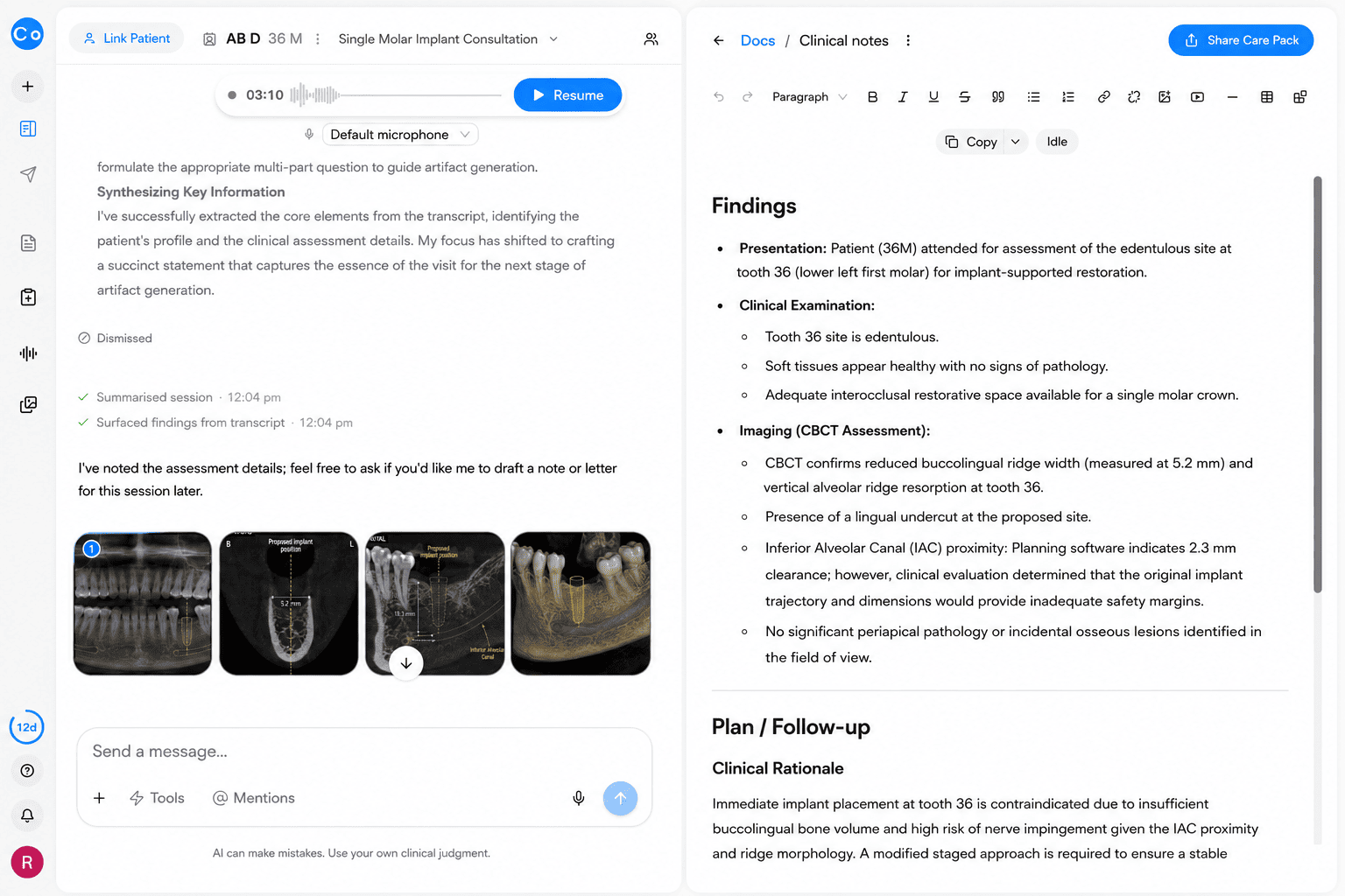
CoTreat Chairside clinical note interface showing an AI-generated Findings document for a single molar implant consultation at tooth 36, recording CBCT-confirmed reduced buccolingual ridge width (5.2 mm), vertical ridge resorption, a lingual undercut, and inferior alveolar canal clearance of 2.3 mm judged an inadequate safety margin for the original implant trajectory, with no periapical pathology or incidental findings. Demonstrates AI-assisted structured clinical documentation of CBCT findings for implant treatment planning.
Enlarge
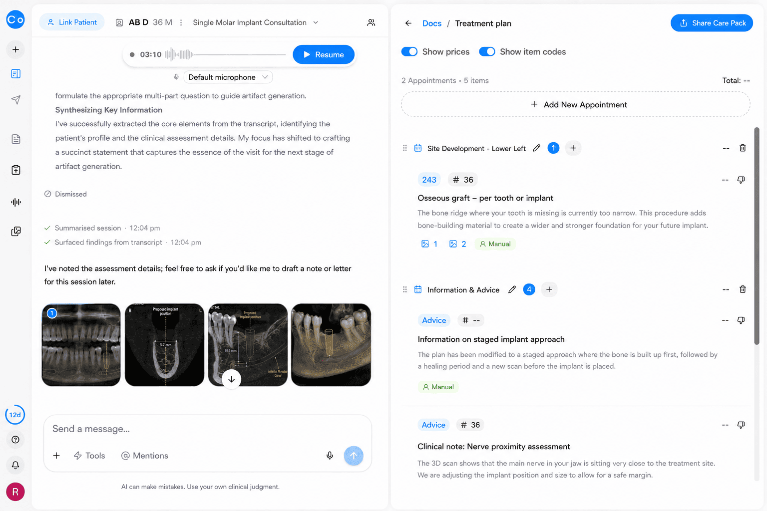
CoTreat Chairside treatment plan interface displaying AI-generated advice items for an implant case, including a site-development appointment for an osseous graft to widen the deficient ridge, a note on the staged implant approach where the graft heals before a new scan and implant placement, and a clinical note flagging inferior alveolar nerve proximity requiring an adjusted implant position and size. Demonstrates how CBCT findings are translated into a documented, staged implant treatment plan.
Enlarge

Clinical Documentation Framework

What should every structured clinical record contain?

Clinical Documentation

Record:

  • The clinical question that prompted CBCT
  • Bone height, width, and angulation
  • Ridge morphology and undercuts
  • Anatomical structures (canal, foramen, sinus, nasopalatine canal, adjacent roots)
  • Dehiscence and fenestration risk
  • Existing pathology
  • Incidental findings
  • How findings influenced the treatment plan

Clinical Reasoning

A recommendation is only as defensible as the findings that support it. If the decisive CBCT findings are missing from the record, the decision becomes difficult to justify later.

See this workflow in CoTreat Chairside

Explore how consultation, documentation and treatment planning are generated in a real clinical workflow.

Clinical Pearl

Good documentation is not simply a list of measurements.

Well-documented CBCT interpretation is often more valuable than a detailed list of measurements. The record should clearly explain how the findings led to the final treatment recommendation.

πŸ’¬ Patient Communication

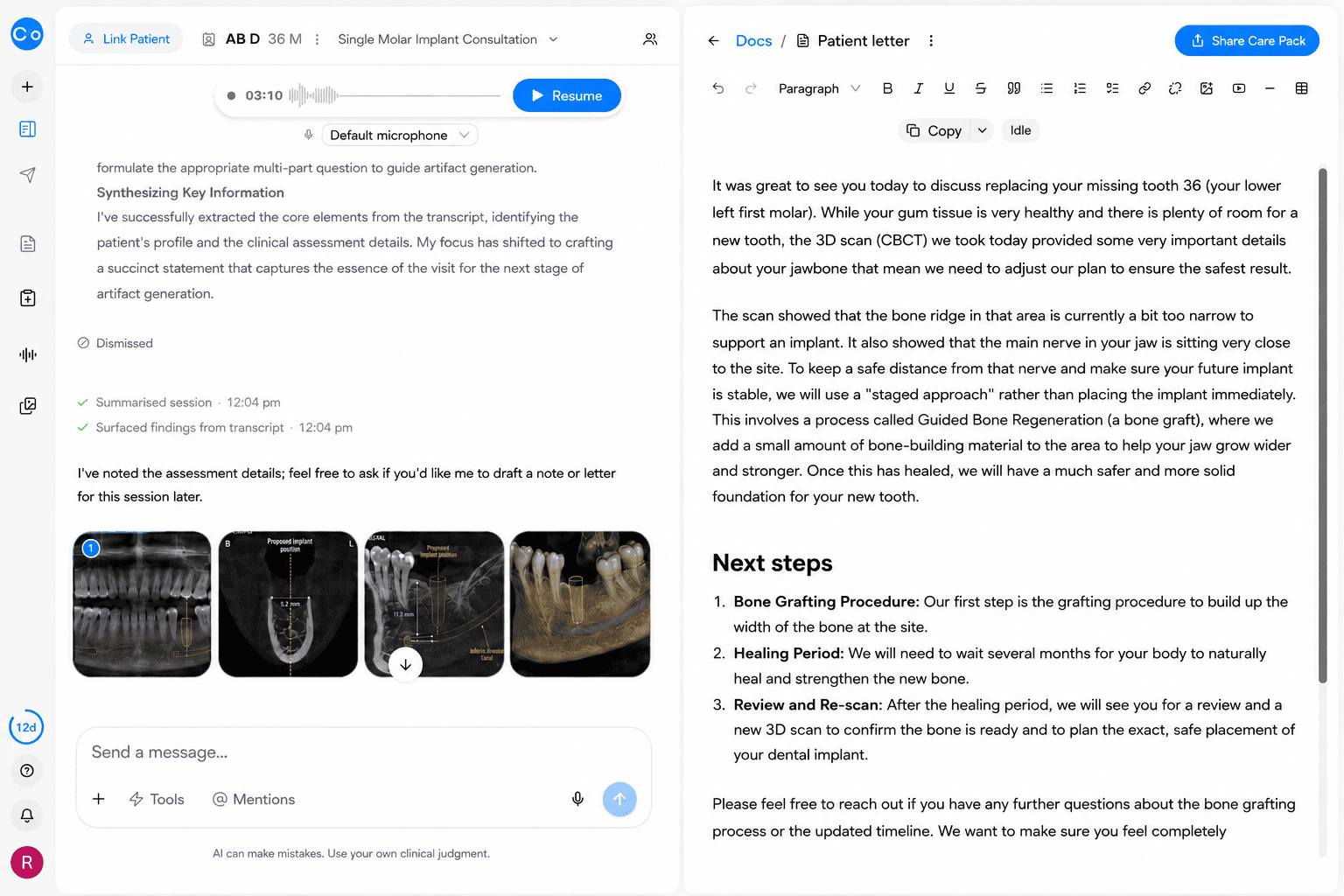
CoTreat Chairside patient letter editor showing an AI-generated letter for an implant consultation explaining, in plain language, why the CBCT scan of the missing lower left first molar site showed a bone ridge that is too narrow and a nerve positioned close to the site, requiring a staged approach of bone grafting, a healing period, and a new scan before implant placement. Demonstrates AI-assisted patient communication translating CBCT findings into understandable language.
Enlarge

Patients should understand why imaging findings require modification, additional treatment, or referral.

How Should CBCT Findings Be Explained?

Communication should support informed, shared decision-making. Patients should understand why CBCT was needed, what it showed, and how the findings changed the implant plan.
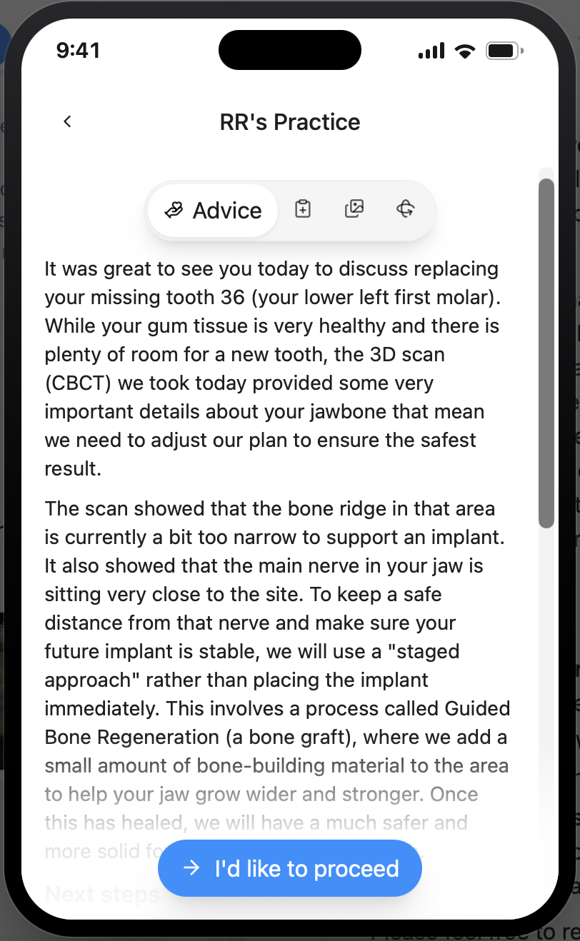
CoTreat Patient Care Pack mobile interface showing the patient-facing version of an implant consultation letter, explaining in plain language that the 3D scan showed a narrow bone ridge and a nearby nerve requiring a staged bone-grafting approach, alongside a button for the patient to indicate they would like to proceed. Demonstrates patient-friendly, mobile communication of CBCT findings and next steps.
Enlarge
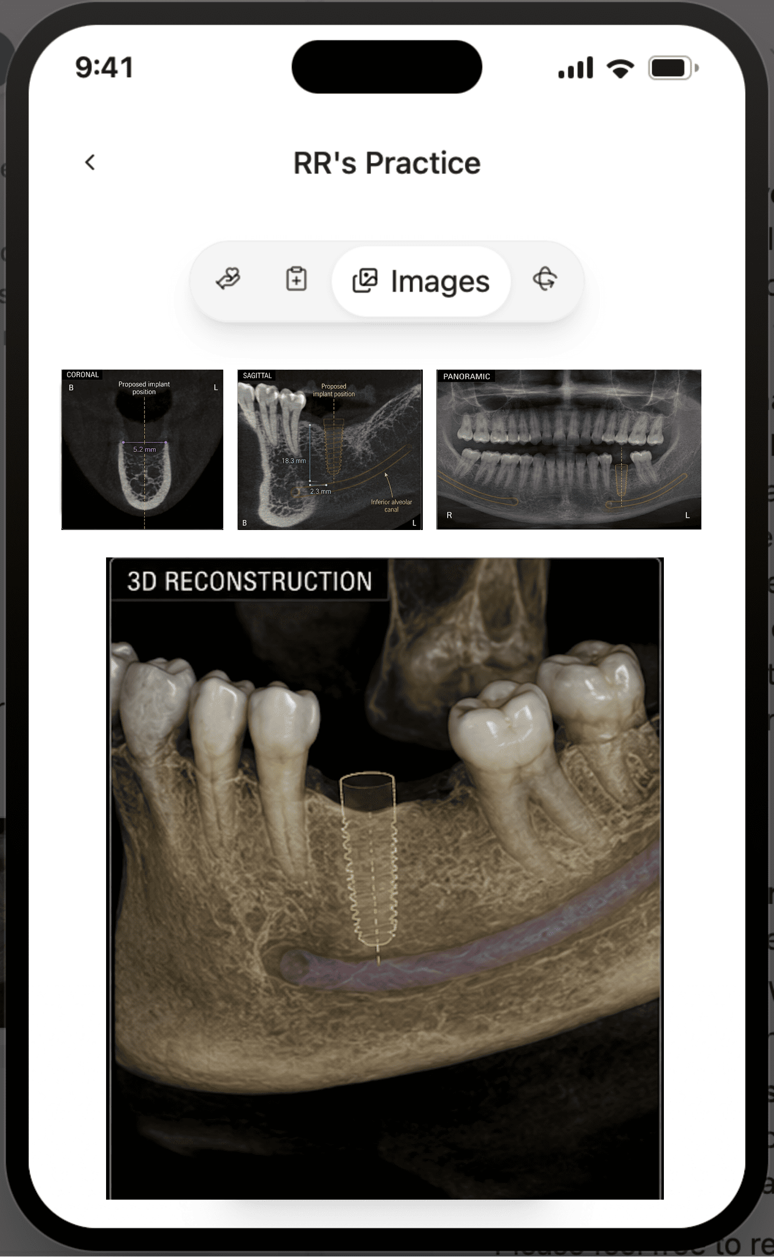
CoTreat Patient Care Pack mobile interface Images tab showing the patient's own CBCT coronal, sagittal, and panoramic views alongside a 3D reconstruction with the inferior alveolar canal highlighted in purple beneath the proposed implant position. Demonstrates giving patients direct, understandable access to their own CBCT imaging alongside the consultation letter.
Enlarge

Patient Communication Framework

How can CBCT findings be translated into patient understanding?

See this workflow in CoTreat Chairside

Explore how consultation, documentation and treatment planning are generated in a real clinical workflow.

Clinical Pearl

Good communication does not oversimplify what the scan showed.

Clear communication builds realistic expectations, supports informed consent, and improves long-term adherence to maintenance, whether the plan proceeds as designed, is modified, staged, or referred.

🌍 CoDL Learning

CBCT interpretation is a critical step in the implant treatment pathway.

Continue Learning

Explore related topics to support treatment planning, site development, complications, and long-term maintenance.

Learning Pathways

Clinical Pearl

CBCT interpretation is the gateway to safe and predictable implant treatment.

A structured assessment provides the foundation for planning, surgery, restoration, maintenance, and long-term clinical success.


About CoTreat

CoTreat is building the AI colleague for dentists.

CoTreat makes it easy to go above and beyond for your patient. An AI that supports you in clinic.