Clinical Reference for Dental Professionals

When Should I Choose Immediate, Early, or Late Implant Placement?

Determine whether immediate, early, or late implant placement gives the most predictable outcome for this site.

๐Ÿงญ Clinical Decision Snapshot

The relevant question is not "Can an implant be placed now?" but "Can this site support predictable, restoratively driven implant placement now?"

Implant timing should be determined by asking whether the site can support predictable, restoratively driven implant placement now, or whether healing, site development, risk modification, or referral will make the outcome more predictable. Immediate placement should be considered only when the clinician has the surgical and restorative experience required for predictable execution.

Implant timing should be considered together with site management; changing timing does not replace the need to manage deficient bone, soft tissue, infection, or other local risk factors.

Key Clinical Takeaways
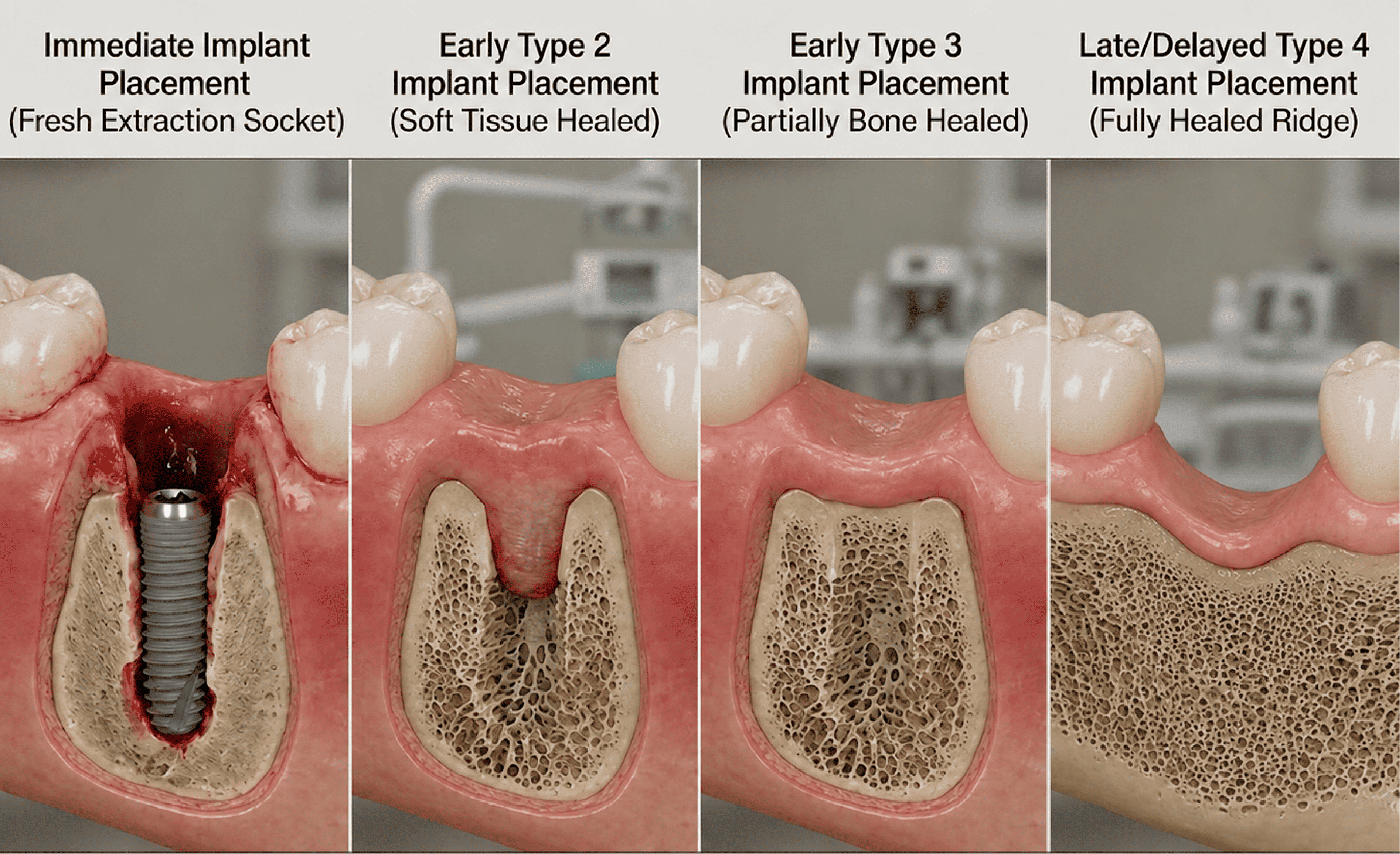
Four-panel cross-sectional comparison of implant placement timing: immediate placement into a fresh extraction socket, early Type 2 placement after soft-tissue healing, early Type 3 placement after partial bone healing, and late/delayed Type 4 placement into a fully healed ridge. Demonstrates the progressive bone fill and soft-tissue closure across the four implant timing pathways.
Enlarge

๐Ÿ” Clinical Assessment

Evaluate socket-wall integrity, infection, soft tissue, primary stability, aesthetic risk, and patient factors.

Which Factors Should Determine Implant Timing?

Implant timing should be determined by integrating socket-wall integrity, infection status, soft-tissue phenotype, primary stability in restoratively driven position, aesthetic risk, CBCT/anatomic assessment, and patient-related factors.

Clinical Assessment Framework
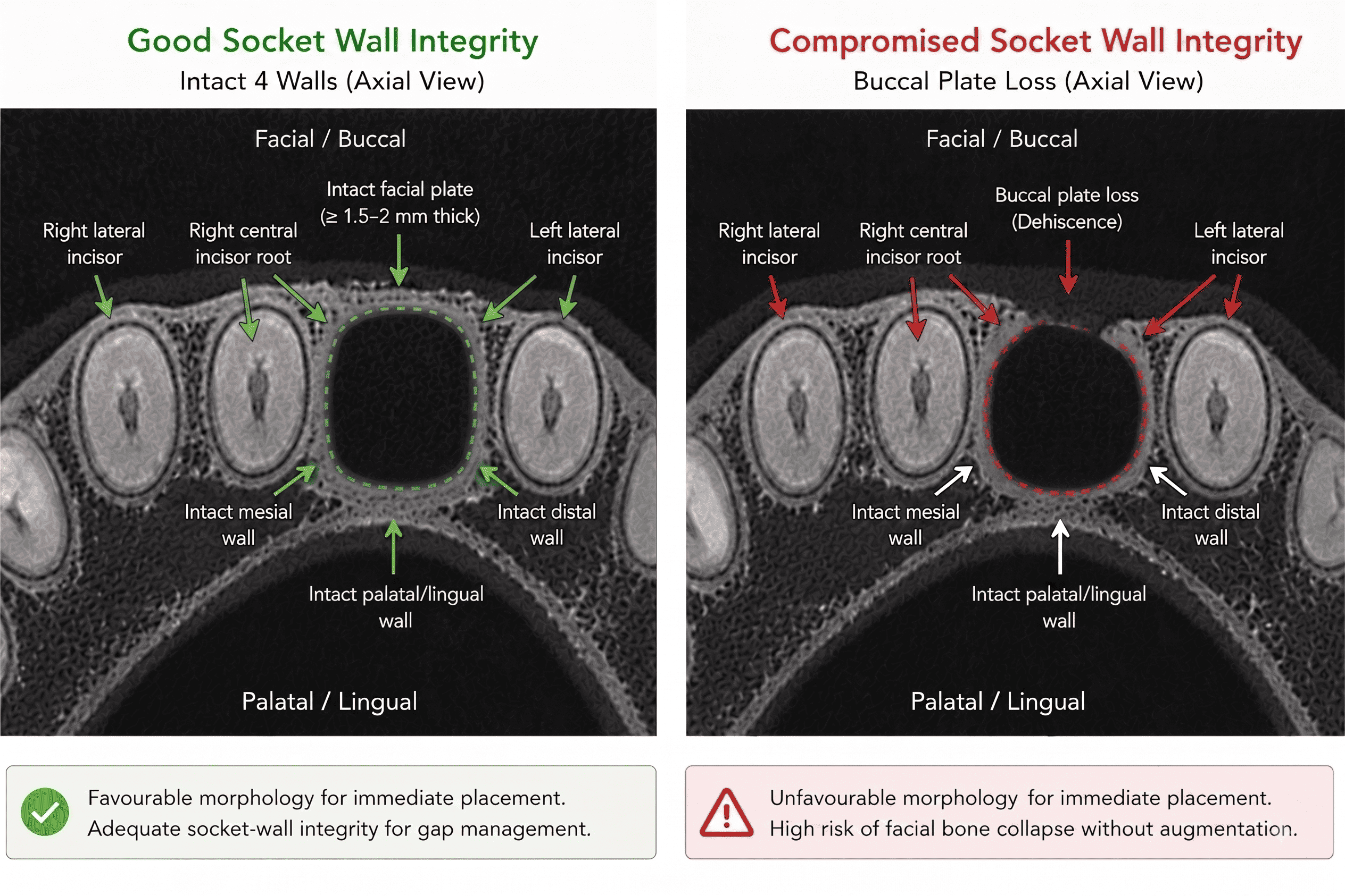
Side-by-side axial CBCT comparison labelled Good Socket Wall Integrity, showing an intact facial plate at least 1.5 to 2mm thick with intact mesial, distal, and palatal/lingual walls, versus Compromised Socket Wall Integrity, showing buccal plate loss (dehiscence) with the other three walls still intact. Demonstrates how socket-wall morphology determines favourability for immediate implant placement.
Enlarge
Socket-Wall Integrity

Clinical Assessment

Evaluate:

  • Intact socket walls
  • Buccal/facial plate integrity and thickness (1.5 to 2mm preferred)
  • Presence of dehiscence or fenestration
  • Defect morphology (3- or 4-wall defect preferred for gap grafting)
  • Ability to achieve appropriate implant positioning and primary stability
  • Facial/buccal bone wall integrity in the aesthetic zone
  • Tooth position within the facial-palatal/lingual bone envelope
  • Gap width between implant and facial plate

Clinical Reasoning

In Type 1 placement, a buccal gap greater than 2mm should be filled with a low-substitution bone graft (e.g., DBBM) to prevent long-term facial bone collapse, even when the facial plate is intact. A 3- or 4-wall defect is typically required to contain gap grafting materials without a full barrier membrane.

A common pitfall is attempting immediate placement in sockets with significant buccal plate defects or unfavourable morphology without considering augmentation options, or failing to graft the buccal gap in Type 1 cases.

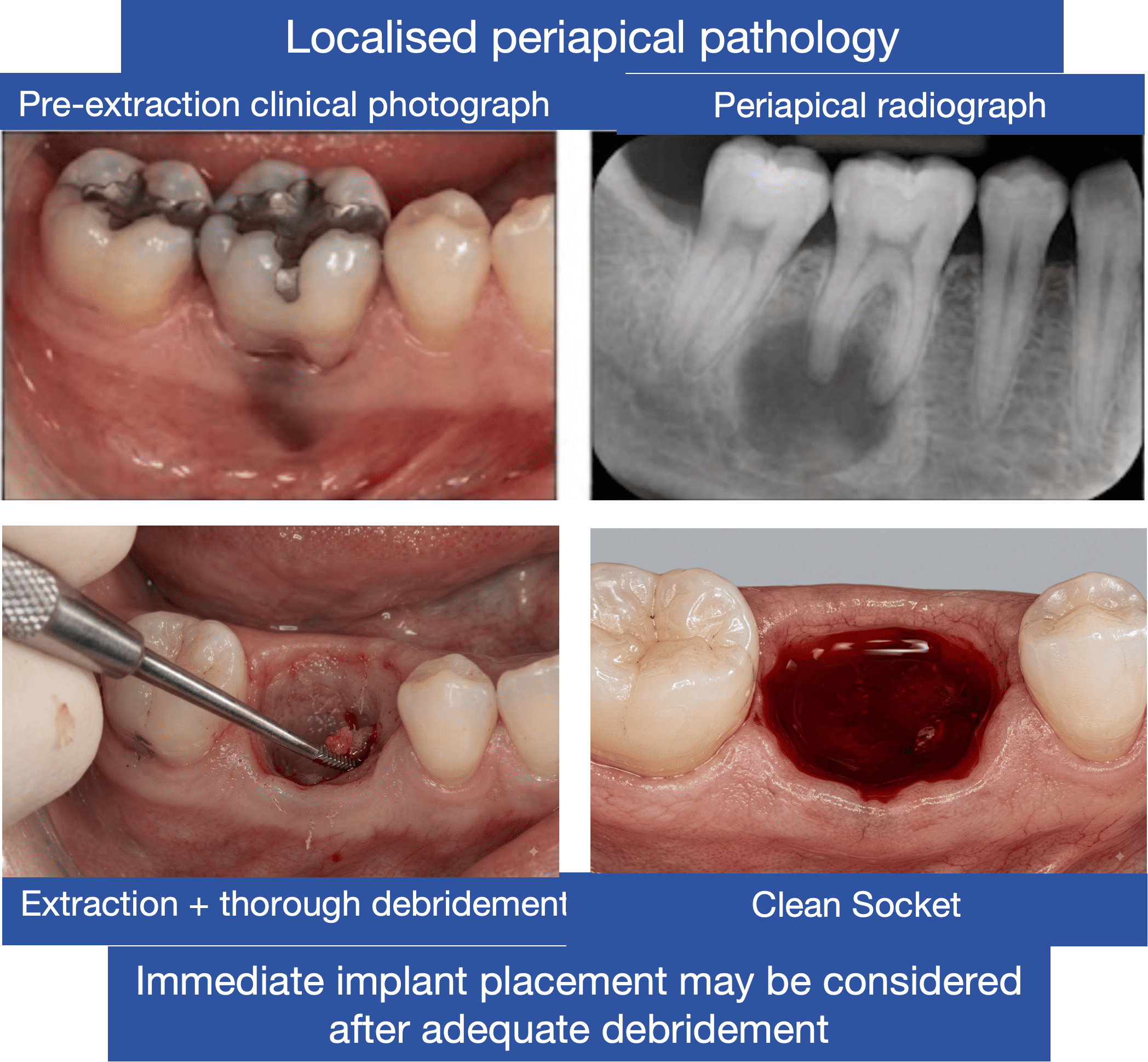
Four-panel clinical case labelled Localised Periapical Pathology, showing a pre-extraction clinical photograph, a periapical radiograph, extraction with thorough debridement, and the resulting clean socket. Demonstrates that chronic, localised periapical pathology may still permit immediate implant placement after adequate debridement.
Enlarge
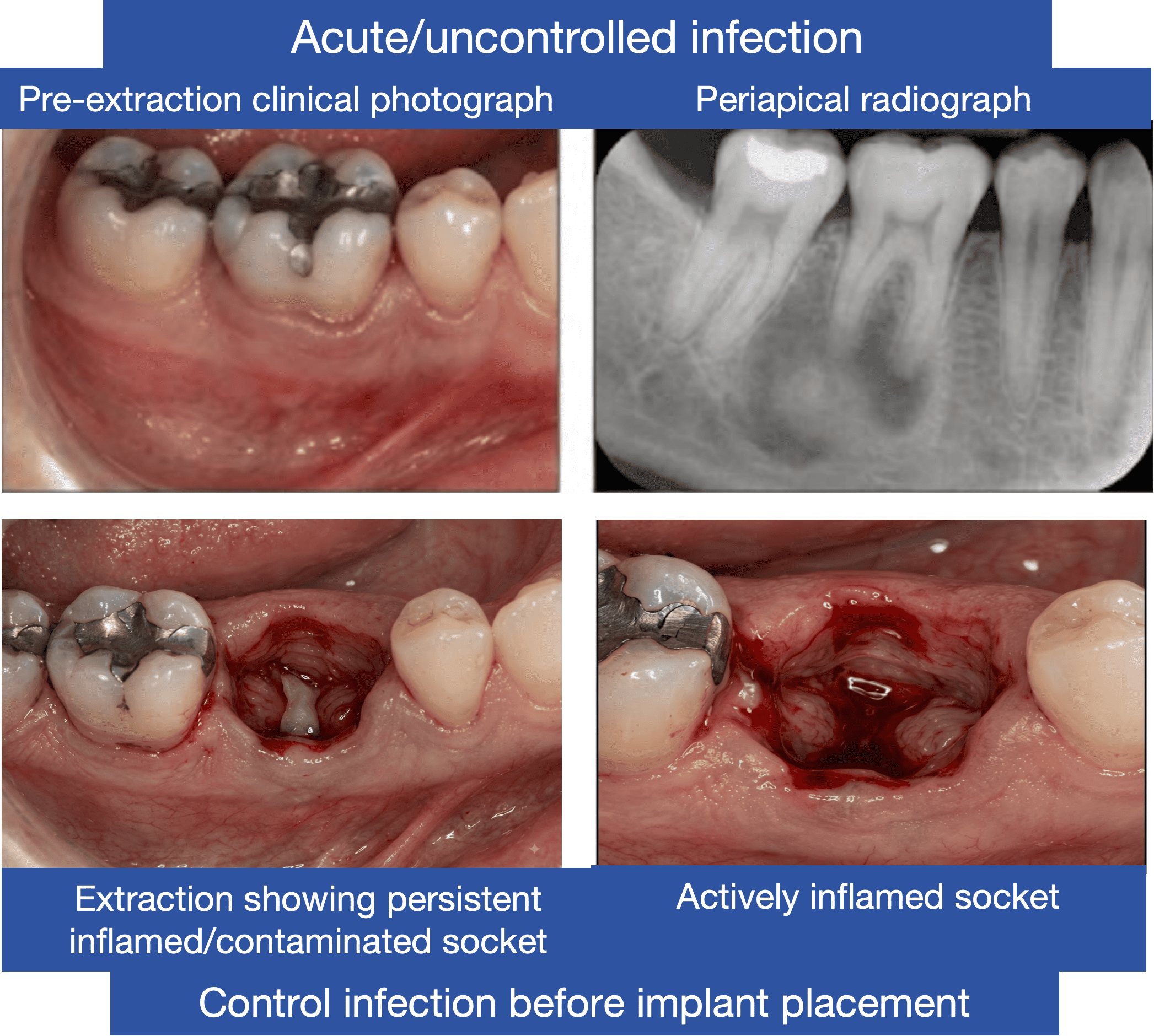
Four-panel clinical case labelled Acute/Uncontrolled Infection, showing a pre-extraction clinical photograph, a periapical radiograph, extraction showing a persistent inflamed and contaminated socket, and an actively inflamed socket after extraction. Demonstrates that acute or uncontrolled infection should be controlled before implant placement.
Enlarge
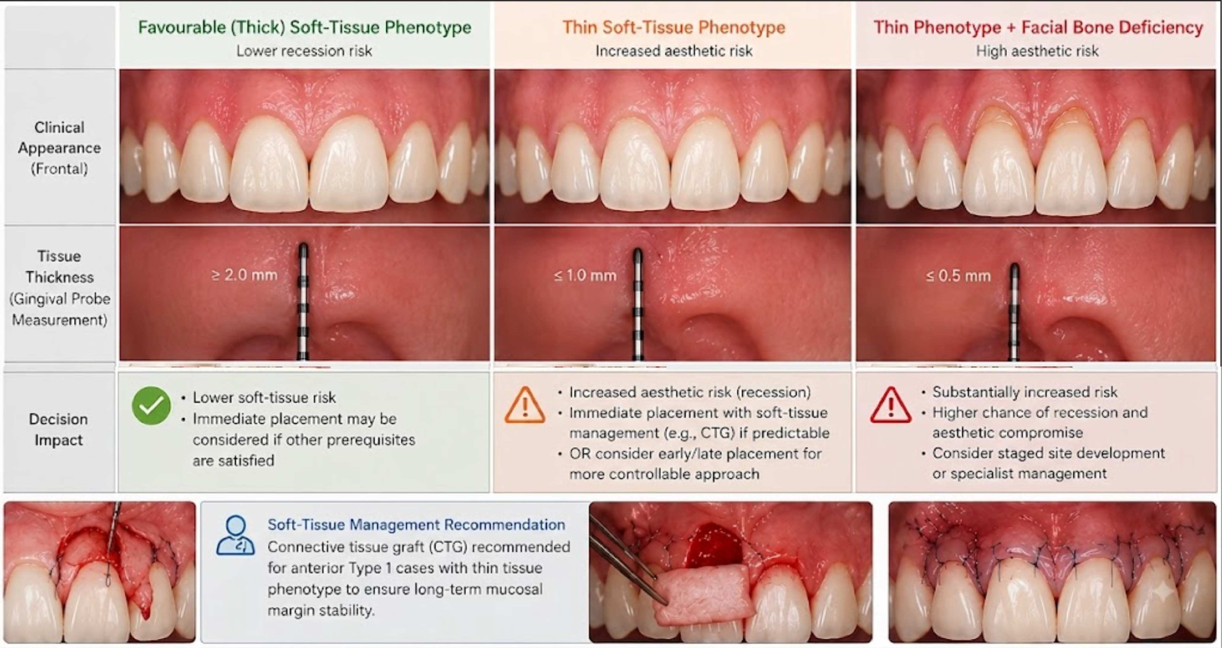
Three-column comparison of favourable thick soft-tissue phenotype (tissue thickness at least 2.0mm, lower recession risk), thin soft-tissue phenotype (1.0mm or less, increased aesthetic risk), and thin phenotype with facial bone deficiency (0.5mm or less, high aesthetic risk), each with a clinical photograph and gingival probe measurement, alongside connective tissue graft (CTG) surgical steps. Demonstrates how measured tissue thickness changes the implant timing and soft-tissue management decision.
Enlarge
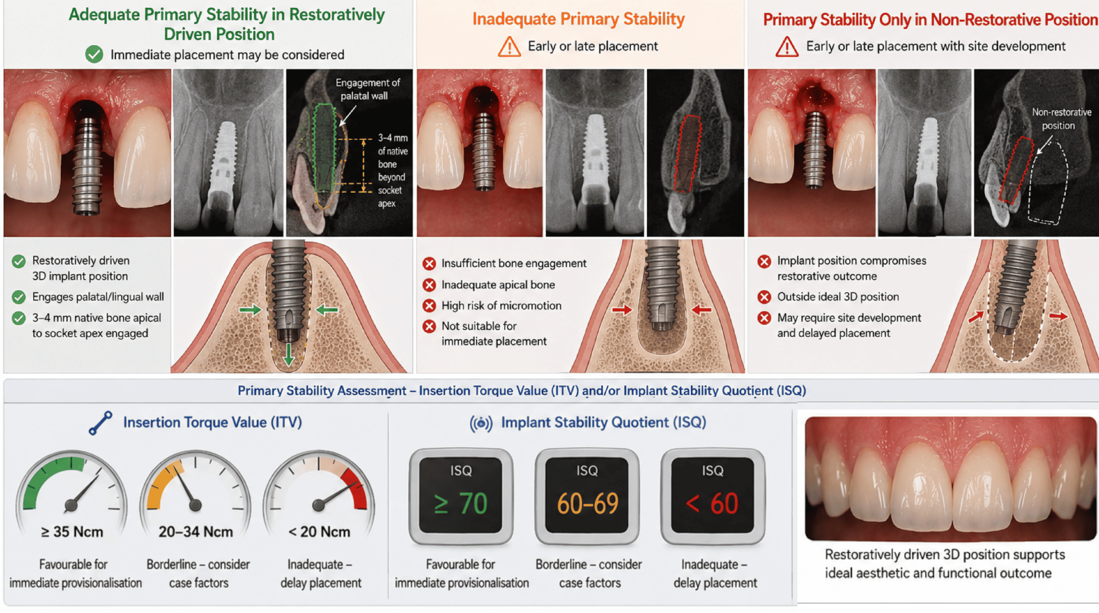
Three-column comparison of adequate primary stability in a restoratively driven position (engaging the palatal wall and 3 to 4mm of native bone apical to the socket), inadequate primary stability with insufficient bone engagement, and stability achieved only in a non-restorative position, alongside insertion torque value (ITV) and implant stability quotient (ISQ) gauges showing favourable, borderline, and inadequate ranges. Demonstrates how primary stability and implant position together determine whether immediate placement or provisionalisation is appropriate.
Enlarge
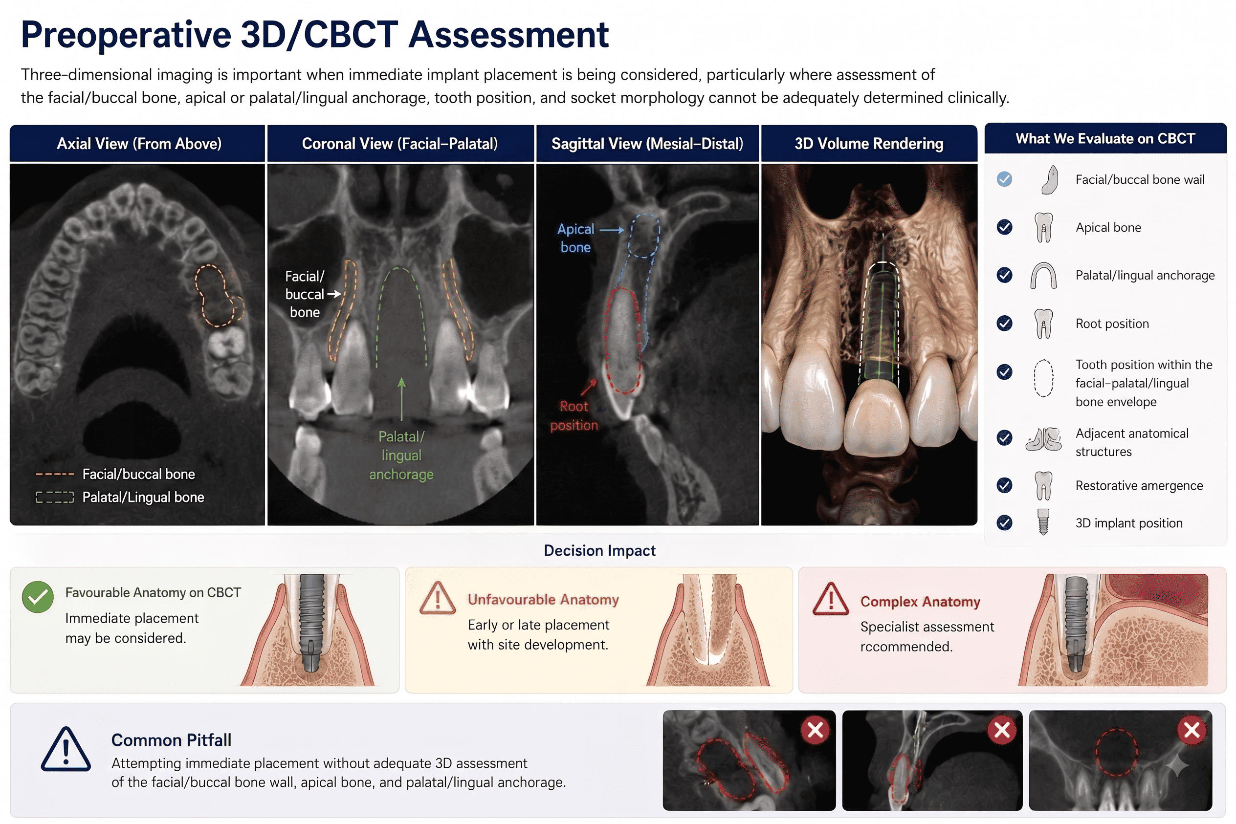
Four-panel CBCT assessment of an anterior extraction site showing axial, coronal, and sagittal views with the facial/buccal bone, palatal/lingual anchorage, apical bone, and root position labelled, alongside a 3D volume rendering of the planned implant, and a checklist of what to evaluate on CBCT including facial/buccal bone wall, apical bone, palatal/lingual anchorage, root position, tooth position within the bone envelope, adjacent structures, restorative emergence, and 3D implant position.
Enlarge
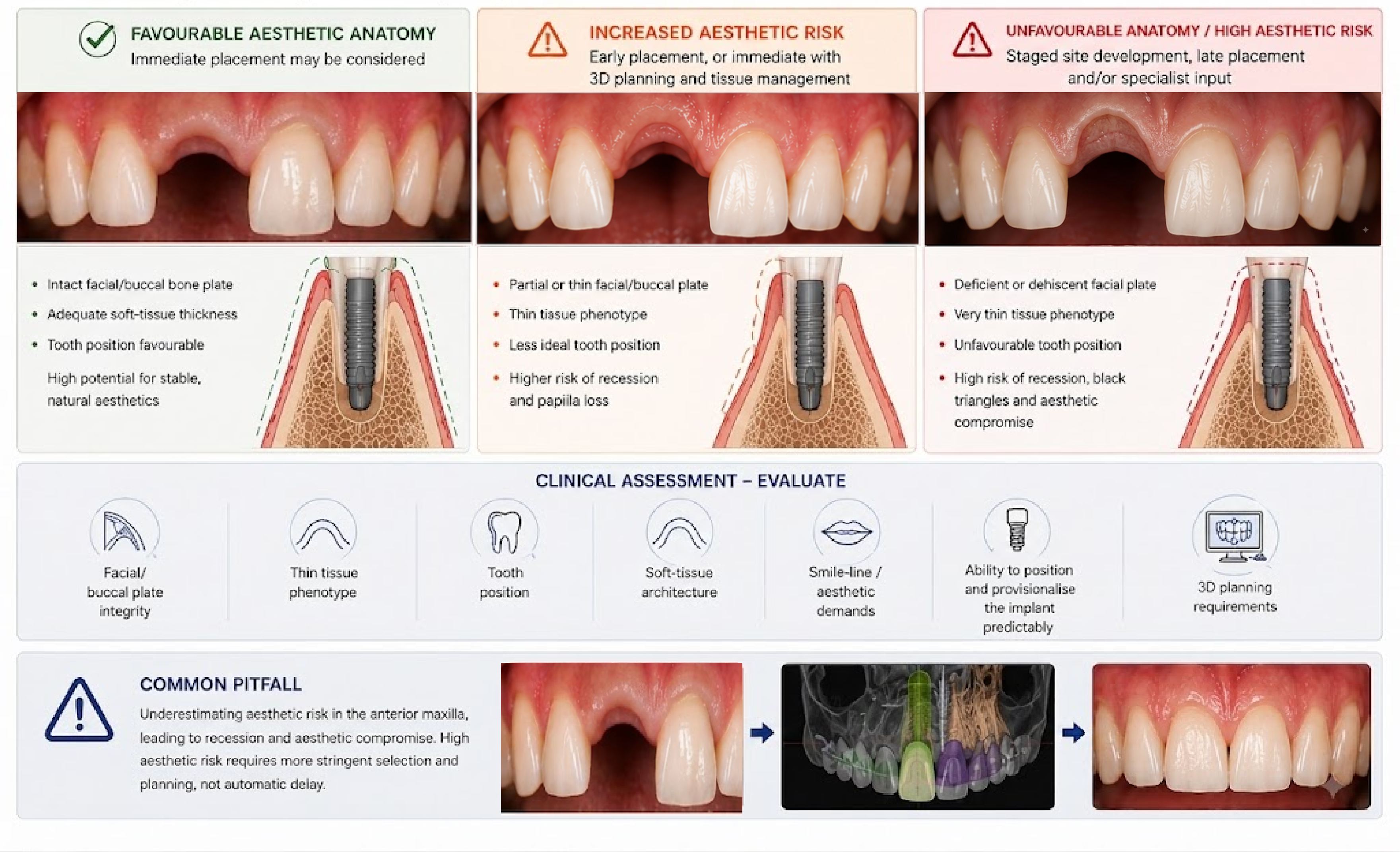
Three-column comparison of favourable aesthetic anatomy (intact facial/buccal bone plate, adequate soft-tissue thickness, favourable tooth position), increased aesthetic risk (partial or thin facial/buccal plate, thin tissue phenotype), and unfavourable anatomy with high aesthetic risk (deficient or dehiscent facial plate, very thin tissue phenotype), each with a clinical photograph and cross-sectional implant diagram, alongside a case sequence from gap to 3D-planned implant to final restoration. Demonstrates how aesthetic-zone risk factors change the timing and planning approach.
Enlarge
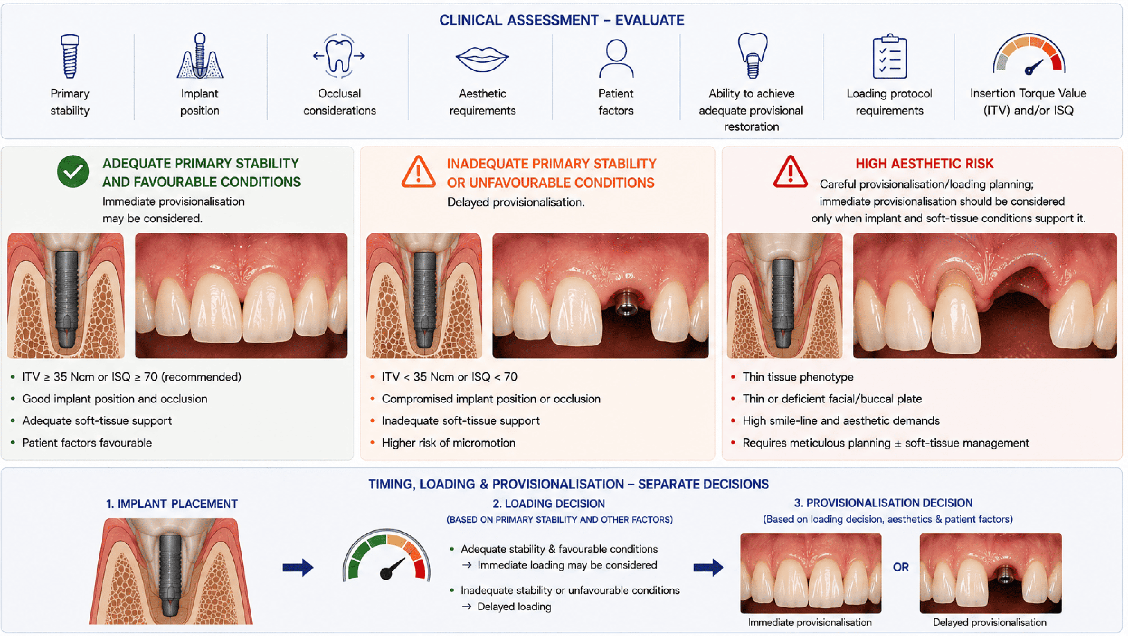
Three-column decision panel showing adequate primary stability and favourable conditions supporting immediate provisionalisation, inadequate primary stability or unfavourable conditions requiring delayed provisionalisation, and high aesthetic risk requiring careful provisionalisation planning, alongside a three-step flow separating implant placement, the loading decision based on primary stability, and the provisionalisation decision based on loading, aesthetics, and patient factors.
Enlarge
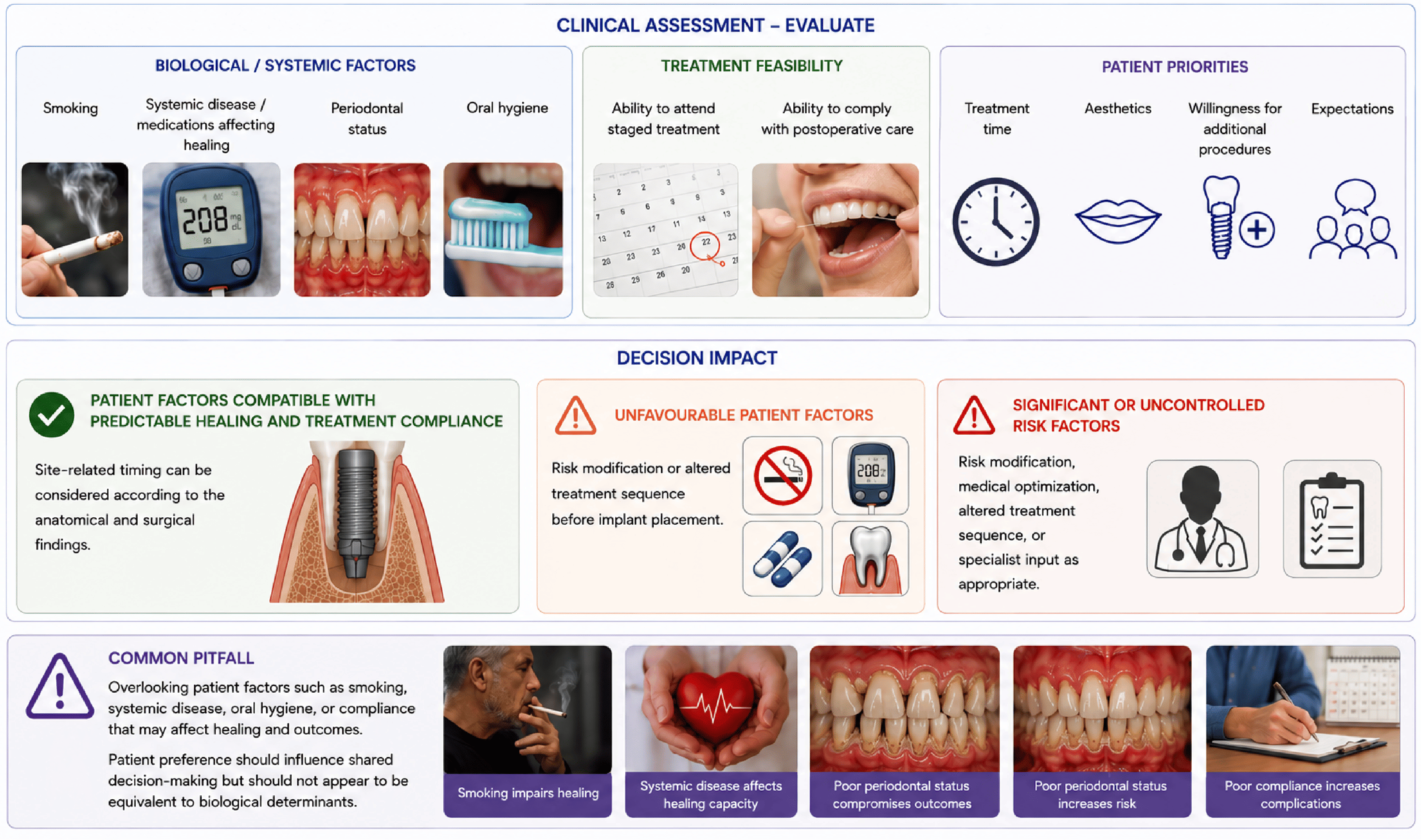
Three-column clinical assessment panel covering biological/systemic factors (smoking, systemic disease or medications, periodontal status, oral hygiene), treatment feasibility (ability to attend staged treatment, ability to comply with postoperative care), and patient priorities (treatment time, aesthetics, willingness for additional procedures, expectations), with a decision-impact row showing favourable, unfavourable, and significant or uncontrolled risk-factor pathways.
Enlarge

Clinical Pearl

Successful implant timing depends on selecting the right pathway for the right patient and site.

Successful implant timing depends on selecting the right pathway for the right patient and site, not simply placing an implant as soon as possible. The critical question is whether the site can support predictable, restoratively driven implant placement now.

โš–๏ธ Clinical Decision-Making

Integrate findings to decide on immediate, early, late placement, or referral.

How Should Implant Timing Be Determined?

Implant timing should be determined by asking whether the site can support predictable, restoratively driven implant placement now, or whether healing, site development, risk modification, or referral will make the outcome more predictable.

Clinical Decision Framework

How do individual findings become one implant timing recommendation?

Immediate (Type 1)

Typical Findings

Typical findings include:

  • Favourable site with intact socket walls
  • No acute or uncontrolled infection
  • Adequate primary stability while maintaining a restoratively driven three-dimensional implant position
  • Manageable aesthetic risk
  • Patient factors compatible with predictable healing and treatment compliance
  • Appropriate clinician experience and surgical capability
  • Gap greater than 2mm managed with bone graft material
  • ITV of at least 35 Ncm or ISQ of at least 70 for immediate provisionalisation

Clinical Reasoning

Implant placement proceeds at the time of extraction.
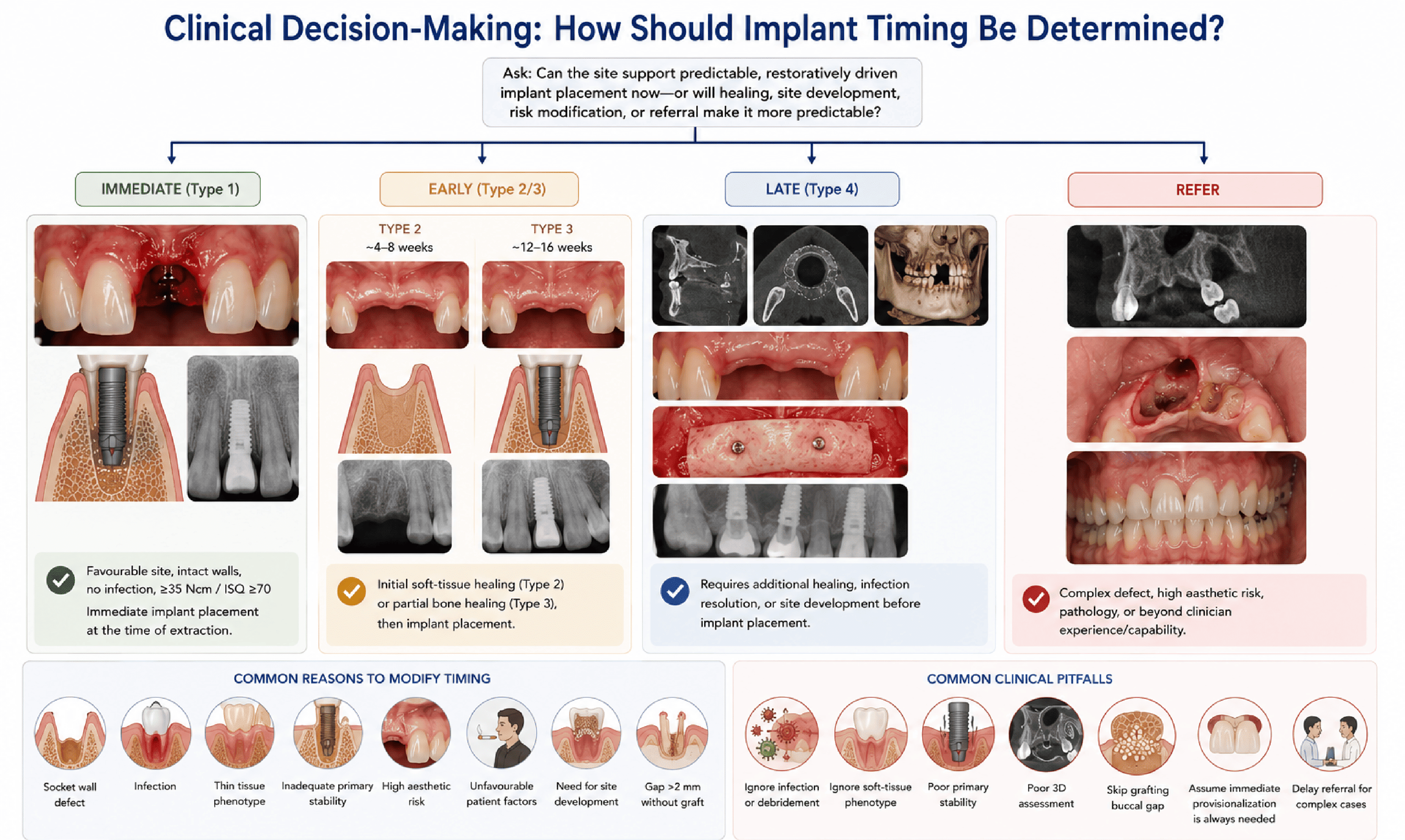
Four-column clinical decision flowchart branching from the question 'Can the site support predictable, restoratively driven implant placement now, or will healing, site development, risk modification, or referral make it more predictable?' into Immediate (Type 1), Early (Type 2/3), Late (Type 4), and Refer pathways, each shown with representative clinical and radiographic images and the typical findings that support that pathway.
Enlarge

Clinical Pearl

The strongest implant timing decisions are made when multiple findings agree.

The strongest implant timing decisions are made when multiple independent findings point toward the same management pathway. When important findings conflict, the safest decision is often to modify timing, delay treatment, or refer rather than proceed immediately.

๐Ÿ“ Clinical Documentation

Document the rationale for timing, risk factors, and treatment plan.

How Should Implant Timing Be Documented?

Clinical records should explain why immediate, early, or late implant placement was recommended. Another clinician should be able to understand the reasoning behind the timing decision.
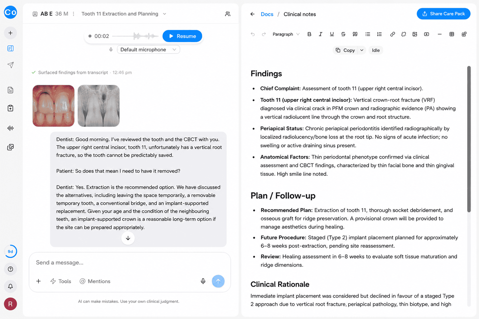
CoTreat Chairside clinical note showing Findings for tooth 11, including a vertical crown-root fracture, chronic periapical periodontitis with no acute infection, and a thin periodontal phenotype with a high smile line, followed by a Plan/Follow-up section recommending extraction with socket debridement, an osseous graft, a provisional crown, and staged Type 2 implant placement at 6 to 8 weeks. Demonstrates structured documentation of the findings and rationale behind a staged implant timing decision.
Enlarge
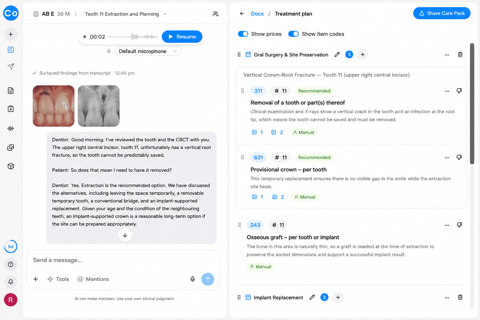
CoTreat Chairside treatment plan showing an Oral Surgery and Site Preservation group with line items for removal of the fractured tooth, a provisional crown, and an osseous graft to preserve the socket, each with a clinical rationale, followed by the start of an Implant Replacement group. Demonstrates how a timing decision is translated into a documented, staged treatment plan.
Enlarge

Clinical Documentation Framework

What should every structured clinical record contain?

Clinical Documentation

Record:

  • Socket-wall integrity and morphology
  • Infection status and debridement
  • Soft-tissue phenotype
  • Primary stability assessment while maintaining a restoratively driven three-dimensional implant position
  • Aesthetic risk evaluation
  • CBCT/anatomic assessment findings
  • Patient factors and preferences
  • Rationale for timing decision
  • Loading/provisionalisation protocol decision
  • Gap management and bone grafting, if applicable
  • ITV/ISQ values, if applicable

Clinical Reasoning

A recommendation is only as defensible as the findings that support it. If the decisive findings are missing from the record, the decision becomes difficult to justify later.

See this workflow in CoTreat Chairside

Explore how consultation, documentation and treatment planning are generated in a real clinical workflow.

Clinical Pearl

Good documentation is not simply a list of findings.

Well-documented clinical reasoning is often more valuable than a detailed list of findings. The record should clearly explain how the assessment led to the final timing recommendation.

๐Ÿ’ฌ Patient Communication

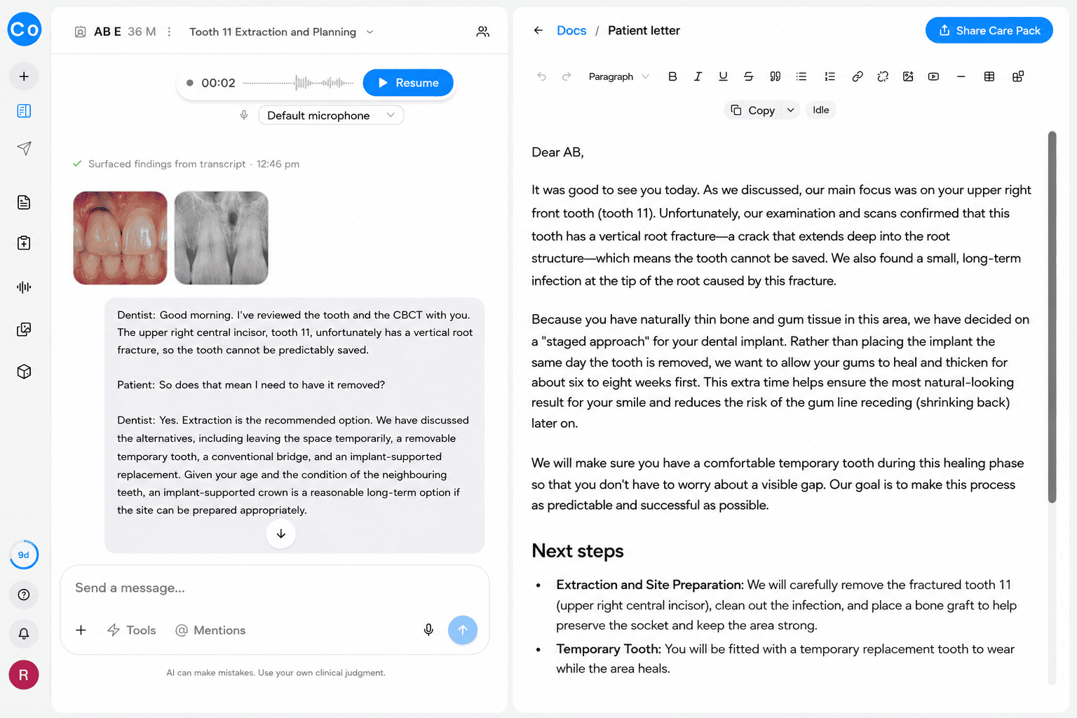
CoTreat Chairside patient letter editor showing an AI-generated letter to a patient explaining, in plain language, that their upper right front tooth has a vertical root fracture and cannot be saved, and that because they have naturally thin bone and gum tissue, a staged approach is recommended: extraction with a bone graft now, six to eight weeks of healing with a temporary tooth, then the dental implant. Demonstrates AI-assisted patient communication translating a timing decision into understandable language.
Enlarge

Patients should understand why immediate, early, or late implant placement is recommended.

How Should Implant Timing Be Explained?

Patients should understand why immediate, early, or late implant placement is recommended, what the risks and benefits are, and what the expected treatment sequence involves.
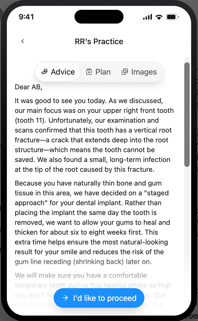
CoTreat Patient Care Pack mobile interface showing the patient-facing version of the consultation letter explaining the vertical root fracture and the staged bone-grafting approach to the implant, alongside a button for the patient to indicate they would like to proceed. Demonstrates patient-friendly, mobile communication of a timing decision and next steps.
Enlarge
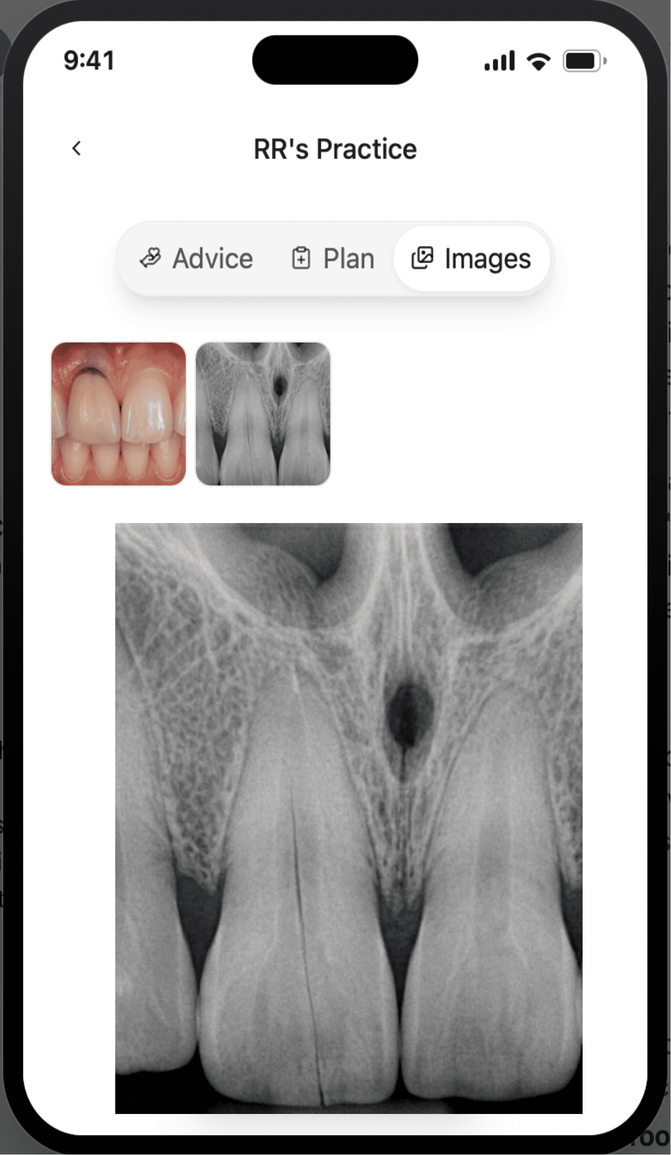
CoTreat Patient Care Pack mobile interface Images tab showing the patient's own clinical photograph and periapical radiograph of the fractured tooth, including an enlarged radiograph view. Demonstrates giving patients direct, understandable access to their own imaging alongside the consultation letter.
Enlarge

Patient Communication Framework

How can a timing decision be translated into patient understanding?

See this workflow in CoTreat Chairside

Explore how consultation, documentation and treatment planning are generated in a real clinical workflow.

Clinical Pearl

Good communication does not oversimplify implant timing.

Clear communication builds realistic expectations, supports informed consent, and improves long-term adherence to maintenance, whether treatment proceeds immediately, is delayed, or referred.

๐ŸŒ CoDL Learning

Implant timing is a critical step in the implant treatment pathway.

Continue Learning

Explore related topics to support socket management, site development, provisionalisation, and long-term maintenance.

Learning Pathways

Clinical Pearl

Implant timing is a critical decision in the implant treatment pathway.

A structured assessment provides the foundation for socket management, surgery, restoration, maintenance, and long-term clinical success.


About CoTreat

CoTreat is building the AI colleague for dentists.

CoTreat makes it easy to go above and beyond for your patient. An AI that supports you in clinic.