Why Does a Root Canal Treated Tooth Become Dark?
“A root canal treated tooth may gradually darken because of internal structural changes, old blood products, restorative materials, or loss of natural translucency.


Short Answer
A root canal treated tooth can become darker over time due to breakdown products inside the tooth, prior pulpal bleeding, internal staining, aging restorations, or changes in how light passes through the tooth after the nerve tissue is removed.
Why Can a Root Canal Treated Tooth Become Darker?
A healthy tooth normally contains:
- living pulp tissue,
- blood supply,
- and natural translucency.
When root canal treatment is performed:
- the pulp tissue is removed,and:
- internal structural and optical changes may gradually affect tooth color over time.
People may notice:
- yellowing,
- gray discoloration,
- darker appearance compared with nearby teeth,
- or gradual color change over months or years.
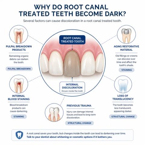
What Commonly Causes Tooth Darkening After Root Canal Treatment?
Darkening may occur because of:
- old blood products from previous inflammation or trauma,
- internal tissue breakdown before treatment,
- staining from older dental materials,
- structural dehydration,
- or altered light-transmission through the tooth.
In some teeth:
- discoloration begins long before treatment,especially when:
- trauma,
- pulpal necrosis,
- or internal bleedingoccurred earlier.
Importantly:discoloration alone does not automatically mean:
- the root canal has failed.
Many darkened teeth:
- remain structurally and biologically stable for years.
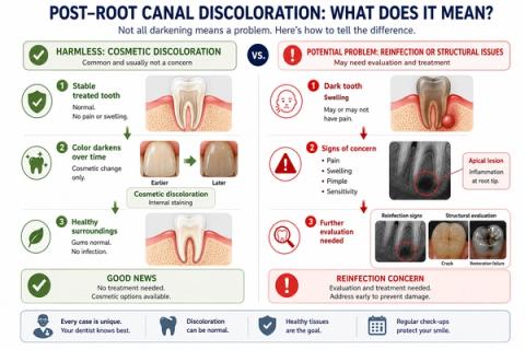
Why the Pattern of Symptoms Matters
| Symptom Pattern | What It May Suggest |
|---|---|
| Darkening without symptoms | Stable cosmetic discoloration |
| Gradual gray or yellow change | Internal staining or optical change |
| Discoloration after trauma | Previous pulpal injury possible |
| Dark tooth with swelling | Possible apical reinfection |
| Pain while chewing or biting | Structural or apical concern |
| Loosening crown or filling | Leakage or restoration failure |
| Progressive symptoms with discoloration | Reassessment needed |
- timing of discoloration,
- radiographic findings,
- restoration condition,
- apical status,
- and structural integrityrather than relying on color alone.
What This Means
The important question is not simply:
“Why is the tooth darker?”
but:
“Does the discoloration represent a cosmetic change alone, or is biologic or structural pathology also present?”
Color changes after root canal treatment often reflect:
- prior biologic injury,
- internal staining,
- restorative material effects,
- and altered dentin light behavior.
However:if discoloration appears together with:
- pain,
- swelling,
- drainage,
- gum changes,
- or biting discomfort,further evaluation may be necessary.
Modern interpretation increasingly focuses on:
- cosmetic stability,
- biologic health,
- restoration integrity,
- and long-term structural prognosis together.
When to See a Dentist
You should consider evaluation if:
- a treated tooth becomes progressively darker,
- swelling develops,
- chewing or biting pain appears,
- gum changes occur near the tooth,
- a crown or filling loosens,
- or discoloration follows trauma.
Color changes may sometimes be cosmetic,but:
- structural and biologic reassessment may still be important.
- apical healing,
- restoration integrity,
- crack risk,
- internal staining causes,
- and long-term structural prognosis—not just the color of the tooth alone.
Early evaluation may help identify:
- cosmetic concerns,
- restorative problems,
- or biologic complicationsbefore they worsen.
Related Questions
Clinical Perspective
For dental professionalsThis section discusses clinical reasoning and is not intended for self-diagnosis.
Post-Endodontic Tooth Discoloration – Pulpal Breakdown and Structural Staining
Clinical Takeaway
Darkening of root canal treated teeth commonly reflects:
- internal chromogenic changes from pulpal degradation,
- hemorrhagic byproducts,
- restorative materials,
- or altered optical propertiesrather than:
- active reinfection alone.
Interpretation Framework
Post-endodontic discoloration should be interpreted as an interaction between:
- pulpal tissue degradation,
- restorative history,
- structural optical changes,
- prior trauma,
- and biologic stability.
Clinical assessment requires integration of:
- discoloration timing,
- treatment history,
- restoration materials,
- vitality status of adjacent teeth,
- apical radiographic findings,
- structural integrity,
- and symptom presence.
The key challenge is distinguishing:
stable cosmetic discoloration
from:
discoloration associated with persistent pathology or structural compromise.
Current interpretation increasingly emphasizes:
- biologic context,
- restorative aging behavior,
- and optical-structural analysis.
Current Understanding (Guidelines + Evidence)
Restorative / Endodontic Perspective
Common causes of post-RCT discoloration include:
- intrapulpal hemorrhage products,
- necrotic tissue remnants,
- sealer or restorative material staining,
- trauma-related pulpal degeneration,
- and altered dentin translucency.
Important interpretation principles include:
- discoloration alone poorly predicts endodontic failure,
- traumatic injuries frequently precede delayed discoloration,
- older endodontic materials historically contributed to staining,
- and structural dehydration and altered light scattering influence appearance.
Biologic Insight
Hemoglobin degradation products may:
- penetrate dentinal tubules,
- alter chromogenic behavior,
- and create long-term intrinsic staining.
Dentin optical behavior changes following:
- pulpal loss,
- structural aging,
- and restorative alteration.
Chronic inflammatory or traumatic events may alter:
- internal pigmentation,
- translucency,
- and dentin reflectivitylong before treatment occurs.
Differential Diagnosis
1. Stable Post-Endodontic Discoloration
Features:
- asymptomatic,
- stable radiographic appearance,
- cosmetic concern primarily.
2. Trauma-Associated Discoloration
Features:
- prior injury history,
- delayed pulpal necrosis possible,
- internal blood-product staining.
3. Persistent/Recurrent Endodontic Pathology
Features:
- associated symptoms,
- radiographic lesions,
- reinfection concern,
- biologic reassessment indicated.
4. Restorative Material Staining
Features:
- localized discoloration patterns,
- older sealers or metallic materials,
- structurally stable tooth.
Key Diagnostic Distinctions
| Feature | Stable Discoloration | Pathology-Associated Concern |
|---|---|---|
| Symptoms | Usually absent | Possible pain/swelling |
| Radiographic findings | Stable | Lesion/reinfection possible |
| Progression | Often gradual/stable | Progressive with other findings |
| Structural integrity | Preserved | May be compromised |
| Cosmetic impact | Primary issue | Secondary to biologic concern |
Common Pitfalls
Common diagnostic errors include:
- assuming all darkened teeth are reinfected,
- missing trauma-related pulpal history,
- overlooking vertical fracture risk,
- ignoring associated apical findings,
- and confusing restorative staining with biologic pathology.
Interpretation should always integrate:
- structural stability,
- biologic status,
- and restorative history.
Emerging Research Directions
Tooth-Color Analytics
Research increasingly focuses on:
- optical translucency modeling,
- internal chromogenic characterization,
- minimally invasive whitening approaches,
- and dentin-light interaction behavior.
AI-Assisted Interpretation
Emerging systems increasingly evaluate:
- discoloration-pattern classification,
- reinfection-risk prediction,
- structural compromise analytics,
- and cosmetic-versus-biologic differentiation. [Schwendicke F et al.]
Advanced Imaging
Current research increasingly explores:
- internal defect visualization,
- restorative leakage assessment,
- structural fatigue mapping,
- and optical-analysis integration.
AI Potential
Darkening of root canal treated teeth represents a:
- biologic-versus-cosmetic interpretation problemwhere:
- optical changes,
- restorative history,
- and structural integritymust be assessed together.
AI can assist across the clinical workflow:
Interpretation
- Integrating discoloration patterns, imaging, restoration status, and symptom history
- Identifying clinically meaningful cosmetic versus pathology-associated changes
Decision Timing
- Supporting monitoring versus retreatment evaluation
- Flagging structural-risk or reinfection-risk patterns
- Assisting restorative planning decisions
Patient Communication
- Explaining why treated teeth may darken over time
- Clarifying the difference between cosmetic staining and biologic failure
- Improving understanding of long-term restorative changes
Clinical Workflow Support
- Structuring discoloration assessment consistently
- Supporting longitudinal monitoring
- Reducing variability in cosmetic-versus-biologic interpretation
Emerging Direction
- AI-assisted optical analysis of endodontically treated teeth
- Predictive discoloration-risk modeling
- Integrated cosmetic and biologic outcome analytics
Clinical Relevance
The challenge is not simply identifying discoloration — it is determining whether the color change reflects:
- stable structural aging,
- prior biologic events,
- restorative material effects,
- or active pathology requiring intervention.
AI may eventually help:
- improve interpretation of post-endodontic discoloration,
- support earlier recognition of pathology-associated changes,
- reduce variability in retreatment assessment,
- and enhance patient communication regarding cosmetic versus biologic concerns.
References
- American Association of Endodontists (AAE). Considerations for the Restoration of Endodontically Treated Teeth. AAE Clinical Resources.
- Plotino G, Buono L, Grande NM, Pameijer CH, Somma F. Nonvital tooth bleaching: a review of the literature and clinical procedures. Journal of Endodontics.
- Lenherr P, Allgayer N, Weiger R, Filippi A, Attin T, Krastl G. Tooth discoloration induced by endodontic materials: a laboratory study. International Endodontic Journal.
- Abbott PV, Heithersay GS, Hume WR. Release and diffusion through human tooth roots in vitro of corticosteroid and tetracycline trace molecules from Ledermix paste. Endodontics & Dental Traumatology.
- Kim ST, Abbott PV, McGinley P. The effects of Ledermix paste on discolouration of mature teeth. International Endodontic Journal.
- Robertson A, Robertson S, Norén JG. A retrospective evaluation of traumatized permanent teeth. International Journal of Paediatric Dentistry.
- Ioannidis K, Mistakidis I, Beltes P, Karagiannis V. Spectrophotometric analysis of coronal discolouration induced by grey and white MTA. International Endodontic Journal.
- Schwendicke F, Samek W, Krois J. Artificial intelligence in dentistry: chances and challenges. Journal of Dental Research.